See the bottom of this page for additional summaries and resources.
Worldwide deployment of 5G, the fifth-generation of cell phone technology, started in 2019. 5G cellular technology employs low-band (600-900 megahertz), mid-band (1.7-4.7 gigahertz), and high-band radio frequencies (24-47 gigahertz).
The allocation of radio frequency spectrum for 5G varies by country. In the United States, the Federal Communications Commission (FCC) has allocated low-band spectrum at 0.6-0.8 GHz (i.e., 600-800 MHz), mid-band spectrum in the 2.5-4.0 GHz range, and 11 GHz of high-band frequencies including licensed spectrum from 24-28 GHz and 37-47 GHz, as well as unlicensed spectrum from 64-71 GHz which is open to all wireless equipment manufacturers.
To increase transmission speed 5G utilizes complex modulation of the carrier wave (i.e., Orthogonal frequency-division multiple access). Other features include massive multiple-input multiple-output (MIMO) or the capacity to send large amounts of data across multiple streams, and beamforming or the use of multiple antennas to control the signal enabling it to be targeted toward specific users. These features can create brief, but very intense, exposures to radio frequency radiation. Since current exposure limits are based upon exposures averaged over time (6 or 30 minutes), these bursts of radiation are essentially unregulated.
Biological and Health Effects of 5G
The 48 studies reported statistically significant evidence of oxidative stress and adverse effects on the neuroendocrine system, the cardiovascular system, sleep quality, sperm quality and DNA damage, bone quality, gene expression, and sensorimotor responses. Most studies used animal models and short-term exposures to microwave radiation (especially continuous wave 3.5 GHz).
However, only eight of these 48 studies actually tested the effects of 5G exposure. The biological and health effects associated with exposure to 5G radiation depend on more than just the carrier frequency. Although these 48 studies employed carrier frequencies common to 5G (e.g., 0.7 GHz, 2.5-3.6 GHz, 27-36 GHz), only eight studies tested exposures with 5G modulation that is likely to have greater impact on health-related outcomes than exposure to continuous waves. Most studies employed a continuous wave generator; yet, 5G requires a complex modulated, orthogonal frequency-division multiplexing (OFDM) signal with additional features (i.e., beamforming, massive MIMO, and phased arrays) that yield brief, high intensity exposures.
Thus, to quote Senator Richard Blumenthal, we are still "flying blind" with regard to ensuring the safety of the population from the short- and long-term effects of widespread exposure to 5G radiation.
The eight 5G studies are briefly summarized below:
Two studies examined the effects of exposure to a 5G cell tower but were subject to confounding with other radio frequency exposures:
- Hardell and Nilsson (2023) reported a case study in which a man and woman developed electromagnetic hypersensitivity (EHS) with neurological symptoms, headache, fatigue, insomnia, tinnitus, skin disorders, and blood pressure variability) after a 5G antenna was added to a 3G/4G cell tower on the roof of their apartment building. (To date, these researchers have published seven case studies with 5G base stations and a summary of all seven reports.)
- Perov et al. (2022) exposed male rats for four months to a 5G base station that transmitted at 3.6 GHz, 28 GHz, and 36 GHz and found that the exposure moderately increased stress on the neuroendocrine system.
Two studies examined the effects of exposure to 5G using 4G/5G cell phones:
- Chu et al. (2023) conducted a pilot study in which human semen samples were briefly exposed to smart phones and found that Wi-Fi negatively affected sperm motility and viability, but not 4G/5G; however, the results varied across phones.
- Pustake et al. (2022) exposed butter bean seeds to a 4G/5G cell phone and found adverse effects on seed germination and growth.
Four studies examined the effects of 5G exposure using a signal generator that simulated 5G base station modulation (but not other features of 5G):
- Krivona et al. (2024) continuously exposed male rats for 4 weeks to 5G radiation (average whole body SAR = 0.0076 W/kg and 0.0059 W/kg; 2.4 GHz carrier frequency) and found no statistically significant cognitive differences in the Morris water maze test as compared to control rats.
- Lameth et al. (2025) exposed adult male mice for 1 hour per day, 5 days per week for 6 weeks to 5G radiation (average brain SAR = 0.19 W/kg, 3.5 GHz carrier frequency) and found mild transcriptome alterations (i.e., gene expression) without changes in memory capacities or emotional state.
- Nikitina et al. (2024) exposed adult Wistar rats to 5G or continuous wave EMF with a frequency of 4.9 GHz and an intensity of 250 μW/cm2 for 15 days for two hours a day and found no differences in open field behavior compared to the control group.
- Sousouri et al (2024) exposed 34 healthy, matched participants in a double-blind, sham-controlled
study with standardized left-hemisphere exposure to two 5G signals (3.6 GHz and 700 MHz) for 30 min before sleep and found that the 3.6 GHz 5G signal modulates spindle center frequency during NREM sleep in a CACNA1C
genotype-dependent manner, implicating LTCC in the physiological
response to RF-EMF and underscoring the need for further research into
5G effects on brain health.
Almost all of the following 48 "5G medical/biological studies" listed in EMF-Portal reported significant adverse biological or health effects:
2025,
Jha N, Sarsaiya P, Tomar AK, Pardhiya S, Nirala JP, Chaturvedi PK, Gupta S, Rajamani P Reprod Toxicol 135: 108910
2025,
Seewooruttun C, Bouguila B, Corona A, Delanaud S, Bodin R, Bach V, Desailloud R, Pelletier A Int J Mol Sci 26 (6): 2792
2025,
Lameth J, Royer J, Martin A, Marie C, Arnaud-Cormos D, Lévêque P, Poirier R, Edeline JM, Mallat M Int J Mol Sci 26 (6): 2459
2025,
Ledent M, Vatovez B, Roelandt P, Bordarie J, Dieudonné M, De Waegeneer E, Kremer C, Boucher L, Bouland C, De Clercq EM Front Public Health 13: 1536167
2025,
Žura N, Vince S, Perić P, Vilić M, Malarić K, Rimac V, Golubić Ćepulić B, Vajdić M, Jurak I, Milinković Tur S, Poljičak Milas N, Samardžija M, Nemir J, Telebuh M, Žura Žaja I Biomedicines 13 (2): 478
2025,
Yilmaz H, Tümkaya L, Mercantepe T, Yilmaz A, Gül F, Suzan ZT Arch Med Res 56 (4): 103157
2024,
Nikitina VN, Kalinina NI, Dubrovskaya EN, Plekhanov VP, Kovshov AA Biol Bull 51 (11): 3473-3480
2024,
Sousouri G, Eicher C, D’ Angelo RM, Billecocq M, Fussinger T, Studler M, Capstick M, Kuster N, Achermann P, Huber R, Landolt HP medRxiv: the Preprint Server for Health Sciences (medRxiv), 2024.12.16.24319082
2024,
Wang X, Zhou G, Lin J, Zhang Z, Qin T, Guo L, Wang H, Huang Z, Ding G Biology 13 (10): 806
2024,
Jamal L, Michelant L, Delanaud S, Hugueville L, Mazet P, Lévêque P, Baz T, Bach V, Selmaoui B Exp Physiol 109 (12): 2122-2133
2024,
Butković I, Vince S, Lojkić M, Folnožić I, Tur SM, Vilić M, Malarić K, Berta V, Samardžija M, Kreszinger M, Žaja IŽ Theriogenology 230: 243-249
2024,
Thoradit T, Chabi M, Aguida B, Baouz S, Stierle V, Pooam M, Tousaints S, Akpovi CD, Ahmad M Commun Integr Biol 17 (1): 2384874
Foroughimehr N, Clayton AHA, Yavari A
Electronics 13 (9): 1630
2024,
Krivova NA, Kudabaeva MS, Zaeva OB, Borodina SV, Lepekhina TB, Pavlenko OA, Makhmanazarov RM, Kokin DS, Shipilov SE Sci Rep 14 (1): 10283
2024,
Zhou GQ, Wang X, Gao P, Qin TZ, Guo L, Zhang ZW, Huang ZF, Lin JJ, Jing YT, Wang HN, Wang CP, Ding GR Sci Total Environ 927: 172391
2024,
Žaja IŽ, Vince S, Butković I, Senaši K, Milas NP, Malarić K, Lojkić M, Folnožić I, Tur SM, Kreszinger M, Samardžija M, Čipčić S, Žura N, Ostović M, Vilić M Animals 14 (6): 828
2024,
Nik Abdull Halim NMH, Mohd Jamili AF, Che Dom N, Abd Rahman NH, Jamal Kareem Z, Dapari R PLoS One 19 (2): e0298738
2024,
Havas F, Cohen M, Krispin S, Attia-Vigneau J Front Biosci (Landmark Ed) 29 (1): 31
2024,
Wang X, Zhou G, Lin J, Qin T, Du J, Guo L, Lai P, Jing Y, Zhang Z, Zhou Y, Ding G Sci Rep 14 (1): 3571
2024,
Torres-Ruiz M, Suárez OJ, López V, Marina P, Sanchis A, Liste I, De Alba M, Ramos V Sci Total Environ: 169475 [in press]
2024,
Handa AP, Vian A, Singh HP, Kohli RK, Kaur S, Batish DR Environ Sci Pollut Res 31 (5): 7465-7480
2024,
Qin TZ, Wang X, Du JZ, Lin JJ, Xue YZ, Guo L, Lai PP, Jing YT, Zhang ZW, Ding GR Int J Environ Health Res 34 (1): 316-327
2023,
Patrignoni L, Hurtier A, Orlacchio R, Joushomme A, Poulletier de Gannes F, Lévêque P, Arnaud-Cormos D, Revzani HR, Mahfouf W, Garenne A, Percherancier Y, Lagroye I Bioelectromagnetics [in press]
2023,
Bodin R, Seewooruttun C, Corona A, Delanaud S, Pelletier A, Villégier AS Environ Sci Pollut Res [in press]
2023,
Jamal L, Yahia-Cherif L, Hugueville L, Mazet P, Lévêque P, Selmaoui B Int J Environ Res Public Health 20 (18): 6793
2023,
Ijima E, Kodera S, Hirata A, Hikage T, Matsumoto A, Ishitake T, Masuda H Front Public Health 11: 1225896
2023,
Canovi A, Orlacchio R, Poulletier de Gannes F, Lévêque P, Arnaud-Cormos D, Lagroye I, Garenne A, Percherancier Y, Lewis N Front Public Health 11: 1231360
2023,
Joushomme A, Orlacchio R, Patrignoni L, Canovi A, Chappe YL, Poulletier de Gannes F, Hurtier A, Garenne A, Lagroye I, Moisan F, Cario M, Lévêque P, Arnaud-Cormos D, Percherancier Y Sci Rep 13: 8305
2023,
Pecoraro R, Pavone SC, Scalisi EM, Ignoto S, Sica C, Indelicato S, Capparucci F, Iaria C, Salvaggio A, Sorbello G, Di Donato L, Brundo MV J Mar Sci Eng 11 (4): 693
2023,
Zheng R, Zhang X, Gao Y, Gao D, Gong W, Zhang C, Dong G, Li Z Brain Behav 13 (6): e3004
2023,
Bektas H, Dasdag S, Nalbant A, Akdag MB, Demir C, Kavak S Biotechnol Biotechnol Equip 37 (1): 2199096
2023,
Qin T, Liu L, Wang X, Guo L, Lin J, Du J, Xue Y, Lai P, Jing Y, Ding G Front Public Health 11: 1087161
2023,
Hardell L, Nilsson M Ann Case Rep 8 (1): 1112
2023,
Chu KY, Khodamoradi K, Blachman-Braun R, Dullea A, Bidhan J, Campbell K, Zizzo J, Israeli J, Kim M, Petrella F, Ibrahim E, Ramasamy R Eur Urol Focus 9 (1): 69-74
2022,
Pustake S, Upadhyaya V, Bundele M 2022 IEEE Pune Section International Conference (PuneCon), Pune, India. IEEE: 1-7, ISBN 978-1-6654-9898-2
2022,
Perov SY, Rubtsova NB, Belaya OV Bull Exp Biol Med 174 (2): 277-279
2022,
Qin TZ, Wang X, Du JZ, Lin JJ, Xue YZ, Guo L, Lai PP, Jing YT, Zhang ZW, Ding GR Int J Environ Health Res [in press]
2022,
Bektas H, Algul S, Altindag F, Yegin K, Akdag MZ, Dasdag S J Chem Neuroanat 126: 102168
2022,
Kim K, Lee YS, Kim N, Choi HD, Lim KM Antioxidants 11 (8): 1449
2022,
Pecoraro R, Pavone SC, Scalisi EM, Sica C, Ignoto S, Contino M, Salvaggio A, Marmara D, Sorbello G, Di Donato L, Brundo MV J Mar Sci Eng 10 (4): 521
2022,
Dasgupta S, Leong C, Simonich MT, Truong L, Liu H, Tanguay RL Environ Sci Technol Lett 9 (4): 327-332
2022,
Yang H, Zhang Y, Wu X, Gan P, Luo X, Zhong S, Zuo W Bioelectromagnetics 43 (2): 106-118
2022,
Wang Y, Jiang Z, Zhang L, Zhang Z, Liao Y, Cai P Environ Pollut 294: 118646
2021,
Wang Y, Zhang H, Zhang Z, Sun B, Tang C, Zhang L, Jiang Z, Ding B, Liao Y, Cai P Environ Pollut 283: 117087
2020,
Kim K, Lee YS, Kim N, Choi HD, Kang DJ, Kim HR, Lim KM Int J Mol Sci 22 (1): E170
2020,
Dasgupta S, Wang G, Simonich MT, Zhang T, Truong L, Liu H, Tanguay RL PLoS One 15 (7): e0235869
A
comprehensive review of 5G NR RF-EMF exposure assessment technologies:
fundamentals, advancements, challenges, niches, and implications
Korkmaz E, Aerts S, Coesoij R, Bhatt CR,
Velghe M, Colussi L, Land D, Petroulakis N, Spirito M, Bolte J. A
comprehensive review of 5G NR RF-EMF exposure assessment technologies:
fundamentals, advancements, challenges, niches, and implications.
Environ Res. 2024 Jul 6;260:119524. doi: 10.1016/j.envres.2024.119524.
Highlights
- Monitoring exposure to radiofrequency electromagnetic fields (RF-EMF) is crucial for environmental health and risk assessment
- A comprehensive review of the diverse landscape of RF-EMF assessment tools was missing.
- There is a definite need for cost-effective and long-lasting EMF sensors.
- Custom-developed RF-EMF measurement tools lack a standardized framework for comparison and validation.
Abstract
This review offers a detailed examination of the current landscape of radio frequency (RF) electromagnetic field (EMF) assessment tools, ranging from spectrum analyzers and broadband field meters to area monitors and custom-built devices. The discussion encompasses both standardized and non-standardized measurement protocols, shedding light on the various methods employed in this domain. Furthermore, the review highlights the prevalent use of mobile apps for characterizing 5G NR radio network data. A growing need for low-cost measurement devices is observed, commonly referred to as “sensors” or “sensor nodes”, that are capable of enduring diverse environmental conditions. These sensors play a crucial role in both microenvironmental surveys and individual exposures, enabling stationary, mobile, and personal exposure assessments based on body-worn sensors, across wider geographical areas. This review revealed a notable need for cost-effective and long-lasting sensors, whether for individual exposure assessments, mobile (vehicle-integrated) measurements, or incorporation into distributed sensor networks. However, there is a lack of comprehensive information on existing custom-developed RF-EMF measurement tools, especially in terms of measuring uncertainty. Additionally, there is a need for real-time, fast-sampling solutions to understand the highly irregular temporal variations EMF distribution in next-generation networks. Given the diversity of tools and methods, a comprehensive comparison is crucial to determine the necessary statistical tools for aggregating the available measurement data.
Conclusions
The
objective of this review was to establish a groundwork for progress in
the field of RF-EMF exposure assessment, ultimately contributing to a
more thorough and efficient assessment. This review provides a
comprehensive overview of the current state-of-the-art concerning RF-EMF
measuring instruments. It covers a wide array of tools, such as
spectrum analyzers, broadband field meters, area monitors, personal
exposimeters, and custom-built instruments, as well as the existing
measurement protocols, encompassing both standardized and
non-standardized methods. In addition, we also have presented some of
the most commonly used mobile apps for collecting 5G NR radio network
data, which have also been used in RF-EMF exposure assessments. However,
it is not yet clear on how accurate the measurement results of these
apps are and how they compare among themselves and to more sophisticated
tools.
Most importantly, this review revealed the
need for cost-effective and long-lasting measurement devices or sensors
that are capable of collecting data at a high time resolution in various
frequency bands, as well as withstanding various environmental
conditions. These sensors are essential for conducting stationary,
mobile, and personal exposure assessments across larger geographical
areas, time intervals, and populations than current capabilities allow.
Additionally, it is important to recognize that the specific
requirements for these sensors differ based on their intended usage,
e.g., on-body measurement devices need to take into account the
influence of the body, vehicle-integrated sensors the influence of the
speed and the relative position of the sensor on the vehicle, and
sensors on infrastructure the influence of the height and the building
materials. Furthermore, there exists a demand for real-time,
fast-sampling solutions to comprehend the highly irregular temporal
variations in EMF distribution within next-generation networks.
Moreover,
there is a notable absence of extensive information regarding currently
employed custom-developed RF-EMF measurement tools, particularly with
respect to measuring uncertainty. Considering the diversity of tools and
methodologies in use, conducting a thorough comparison becomes crucial
to identify the necessary statistical tools for aggregating the
available measurement data.
A more in-depth
discussion relating the current 5G NR assessment methods to measurement
equipment is intended for a follow-up study, which will describe more in
detail the requirements, opportunities, and priorities for new,
low-cost, custom-built measurement equipment.
--
Summary of seven Swedish case reports on the microwave syndrome associated with 5G radiofrequency radiation
Hardell L, Nilsson M. (2024). Summary of seven Swedish case reports on the microwave syndrome associated with 5G radiofrequency radiation. Reviews on Environmental Health. doi: 10.1515/reveh-2024-0017
Abstract
The fifth generation, 5G, for wireless communication is currently deployed in Sweden since 2019/2020, as well as in many other countries. We have previously published seven case reports that include a total of 16 persons aged between 4 and 83 years that developed the microwave syndrome within a short time after being exposed to 5G base stations close to their dwellings. In all cases high radiofrequency (RF) radiation from 4G/5G was measured with a broadband meter. RF radiation reached >2,500,000 to >3,180,000 μW/m2 in peak maximum value in three of the studies. In total 41 different health issues were assessed for each person graded 0 (no complaint) to 10 (worst symptoms). Most prevalent and severe were sleeping difficulty (insomnia, waking night time, early wake-up), headache, fatigue, irritability, concentration problems, loss of immediate memory, emotional distress, depression tendency, anxiety/panic, dysesthesia (unusual touched based sensations), burning and lancinating skin, cardiovascular symptoms (transitory high or irregular pulse), dyspnea, and pain in muscles and joints. Balance disorder and tinnitus were less prevalent. All these symptoms are included in the microwave syndrome. In most cases the symptoms declined and disappeared within a short time period after the studied persons had moved to a place with no 5G. These case histories are classical examples of provocation studies. They reinforce the urgency to inhibit the deployment of 5G until more safety studies have been performed.
--
ICNIRP Guidelines’ Exposure Assessment Method
for 5G Millimetre Wave Radiation May Trigger Adverse Effects
Redmayne M, Maisch DR. ICNIRP Guidelines’ Exposure Assessment Method
for 5G Millimetre Wave Radiation May Trigger Adverse Effects. Int. J. Environ. Res. Public Health 2023, 20, 5267. doi: 10.3390/ijerph20075267.
Abstract
The current global roll-out of 5G infrastructure is designed to utilise
millimetre wave frequencies (30–300 GHz range) at data transmission
rates in the order of gigabits per second (Gbps). This frequency band
will be transmitted using beamforming, a new introduction in near-field
exposures. The International Commission on Non-Ionising Radiation
Protection (ICNIRP) has recently updated their guidelines. We briefly
examine whether the new approach of the ICNIRP is satisfactory to
prevent heat damage and other adverse bio-effects once millimetre wave
5G is included, and we challenge the use of surface-only exposure
assessment for local exposures greater than 6 GHz in part due to
possible Brillouin precursor pulse formation. However, this is relevant
whether or not Brillouin precursors occur from absorption of either 5G
or future G transmissions. Many significant sources conclude there is
insufficient research to assure safety even from the heat perspective.
To date, there has been no published in vivo, in vitro or
epidemiological research using exposures to 5G New Radio beam-formed
signals.
Conclusions
Surface
radiofrequency exposure assessments including mmW radiation are
insufficient to ensure safety; there are several reasons assessment of
SAab is also needed.
A real danger
of the ‘expert’ assurances of a lack of risk is that they discourage the
necessary research to evaluate risk properly. They may also discourage
review of apparently outmoded/questionable approaches being taken in RF
exposure standards.
Once the 5G mmW band is
internationally operational, a significant proportion of the world’s
population will be exposed to new hazards. The intensity and complexity
of near-field exposure, such as when carrying a phone in a pocket or
using it next to the head, will be different for 5G, and this is the
first time mmW have been used for public telecommunications and the
first time beamforming has been deliberately introduced for near-field
use. Without research on the impact of near-field 5G, this global step
is an experiment at the population level. Bearing this in mind, there is
a vital and urgent need for targeted research and for a re-evaluation
of the scientific relevance of the current RF human exposure standards’
basic approach and assumptions.
--
Case
Report: The Microwave Syndrome after Installation of 5G Emphasizes the
Need for Protection from Radiofrequency Radiation
Hardell L, Nilsson M. (2023). Case Report: The Microwave Syndrome after Installation of 5G Emphasizes the Need for Protection from Radiofrequency Radiation. Ann Case Report. 8: 1112. doi: 10.29011/2574-7754.101112.
Abstract
In this case report
two previously healthy persons, a man aged 63 years and a woman aged 62 years, developed symptoms of the microwave syndrome after installation of a 5G base station for wireless communication on the roof above their apartment. A base station for previous telecommunication generation technology (3G/4G) was present at the same spot since several years. Very high radiofrequency (RF) radiation with maximum (highest measured peak value) levels of 354 000, 1 690 000, and >2 500 000 µW/m2 were measured at three occasions in the bedroom located only 5 meters below the new 5G base station, compared to maximum (peak) 9 000 µW/m2 prior to the 5G deployment. The rapidly emerging symptoms after the 5G deployment were typical for the microwave syndrome with e.g., neurological symptoms, tinnitus, fatigue, insomnia, emotional distress, skin disorders, and blood pressure variability. The symptoms were more pronounced in the woman. Due to the severity of symptoms, the couple left their dwelling and moved to a small office room with maximum (peak) RF radiation 3 500 µW/m2. Within a couple of days, most of their symptoms alleviated or disappeared completely. This medical history can be regarded as a classic provocation test. The RF radiation levels in the apartment were well below the limit proposed to be “safe” below which no health effects would occur, recommended by the International Commission on Non-Ionizing Radiation (ICNIRP). These now presented symptoms of the microwave syndrome were caused by non-thermal effects from RF radiation and highlight that the ICNIRP guidelines used in most countries including Sweden do not protect human health. Guidelines based on all biological negative effects from RF radiation are urgently needed, as well as monitoring human health, not the least due to rapidly increasing levels of exposure.
Open access paper:
https://www.gavinpublishers.com/article/view/case-report-the-microwave-syndrome-after-installation-of-5g-emphasizes-the-need-for-protection-from-radiofrequency-radiation
--
Effect of
Radiofrequency Radiation Emitted by Modern Cellphones
on Sperm Motility and Viability: An In Vitro Study
Chu KY, Khodamoradi K, Blachman-Braun R, Dullea A, Bidhan J, Campbell K, Zizzo J, Israeli J, Kim M, Petrella F, Ibrahim E, Ramasamy R. Effect of Radiofrequency Electromagnetic Radiation Emitted by Modern Cellphones on Sperm Motility and Viability: An In Vitro Study. Eur Urol Focus. 2023 Jan;9(1):69-74. doi: 10.1016/j.euf.2022.11.004.
Abstract
Background: Cellphones emit radiofrequency electromagnetic radiation (RF-EMR) for transmission of data for social media communication, web browsing, and music/podcast streaming. Use of Bluetooth ear buds has probably prolonged the time during which cellphones reside in the trouser pockets of men. It has been postulated that RF-EMR increases oxidative stress and induces free radical formation.
Objective: To investigate the effect of wireless-spectrum (4G, 5G, and WiFi) RF-EMR emitted by modern smartphones on sperm motility and viability and explore whether these effects can be mitigated using a physical barrier or distance.
Design, setting, and participants: Semen samples were obtained from fertile normozoospermic men aged 25-35 yr. A current-generation smartphone in talk mode was used as the RF-EMR source. A WhatsApp voice call was made using either 4G, 5G, or WiFi wireless connectivity. We determined if exposure effects were mitigated by either a cellphone case or greater distance from the semen sample.
Outcome measurements and statistical analysis: The semen samples were analyzed according to 2010 World Health Organization laboratory guidelines. Statistical analysis was performed using SPSS v.28.
Results and limitations: We observed decreases in sperm motility and viability with WiFi exposure but not with exposure to 4G or 5G RF-EMR. With large variability among smartphones, continued research on exposure effects is needed.
Conclusions: Our exploratory study revealed that sperm motility and viability are negatively impacted by smartphones that use the WiFi spectrum for data transmission.
Patient summary: We looked at the effect of cellphone use on sperm motility and viability. We found that cellphones using WiFi connectivity for data usage have harmful effects on semen quality in men.
Excerpts
Our study is not without limitations. First, our small sample size of 18
introduces potential sources of bias. We did not collect demographic
data for these patients in order to maintain privacy, so the results may
be subject to confounding bias. As the first of its kind at our
institution, this small trial was a pilot study to validate our
experimental model and procedures. We hope that further studies on the
effects of RF-EMR on semen parameters can be performed on larger samples
to validate our initial results. Second, we recognize that other
potential variables, including temperature and radiation strength, could
play a role in inducing changes in semen parameters. For this
preliminary study, we were only interested in a single variable
(radiation); future work should investigate the impact of temperature
and radiation strength on changes in semen. This was an exploratory in
vitro study, and further in vivo studies in animal models should be
performed to further evaluate the impact of radiation on semen
parameters.
Conclusions
Our
study revealed that 4G/5G RF-EMR emitted by a contemporary cellphone
did not have negative effects on sperm motility and viability. By
contrast, WiFi exposure did have negative effects. During data use,
there may be an increase in heat dissipated by a cellphone, depending on
the power required to connect to the source. Interestingly, we observed
varying effects of WiFi on sperm parameters, depending on the
environment. We posit that a greater distance from the wireless router
results in a need for more cellphone power, which may lead to greater heat production
and result in negative effects on sperm motility and viability.
Mitigation measures such as use of a cellphone case and increasing the
distance between the cellphone and the sperm sample lessened the
effects. Further studies need to be performed to better understand the
effects of RF-EMR on sperm parameters.
--
Status of the Neuroendocrine System in Animals Chronically Exposed to Electromagnetic Fields of 5G Mobile Network Base Stations
Perov SY, Rubtsova NB, Belaya OV. Status of the Neuroendocrine System in Animals Chronically Exposed to Electromagnetic Fields of 5G Mobile Network Base Stations. Bull Exp Biol Med. 2023 Jan 4. doi: 10.1007/s10517-023-05689-2.
Abstract
We studied the biological effect of chronic exposure to multifrequency electromagnetic fields simulating the effects of 5G NR/IMT-2020 mobile communication systems. Male Wistar rats were exposed to 24-h radiation (250 μW/cm2) for 4 months. The exploratory activity of the animals and blood concentrations of ACTH and corticosterone were evaluated at the end of each month of exposure and 1 month after exposure. The results suggest that exposure to multifrequency electromagnetic field simulating the effects of 5G systems affected functional activity of the hypothalamus-pituitary-adrenal axis and was stressful in nature.
Excerpts
The animals were divided into 5 experimental (exposure to EMF of 5G systems, power density (PD) 250 μW/cm2) and 5 control (sham exposure) groups (12 rats each). Exposure conditions: chronic experiment — exposure for 4 months (120 days; 24-h, 7 days per week) and 1-month (30 days) post exposure period (without irradiation). During exposure period, the animals of experimental groups were kept in radio transparent (plastic) cages. Exposure was carried out by 5G/IMT-2020 base stations with simultaneous use of radio channels with 3.6 GHz (n78 with 100 MHz channel bandwidth), 28 GHz (n257 with 100 MHz channel bandwidth) and 37 GHz (n260 with 400 MHz channel bandwidth) central frequencies....
The neuroendocrine system of rats responded to chronic 4-month EMF exposure by waveform changes of serum levels of ACTH and corticosterone. ACTH content had a tendency to increase after 3 months of the experiment (Fig. 1).
Changes in serum corticosterone content in exposed animals were more pronounced; significant differences from the control group were revealed after 1 and 2 months of exposure and the maximum increase was found 1 month after end of exposure (Fig. 2).
Chronic exposure induced changes in orientation and exploratory activity and emotional state of experimental animals. These changes were detected starting from 3rd month of exposure, but did not reach significance threshold, and 1 month after the end of irradiation, the excitation and inhibition processes in the CNS returned to normal.
--
Brillouin
Precursors, a theoretical oddity or a real concern for 5G
millimetre-wave bands to be used in future high-speed
telecommunications?
Don Maisch, Ph.D., Discussion Paper, July 21, 2022
The following topics are briefly discussed in the paper:
- Brillouin Precursors
- The need for reliable research
- Uncertainties with ICNIRP’s thermally based limits for millimeter wave emissions
- A potential risk for property owners
Excerpts
"...
With a millimeter wavelength of 0.65 mm at 42 GHz. The waves can
penetrate into the human skin deep enough to affect most skin structures
located in the epidermis and dermis.1 However, these types of waves
present other challenges. The first is that when most of the energy is
focused in a small area, such as 5G antenna beam-forming, the risk of
human tissue heating for anyone in the path of the beam will be
increased.
The second challenge is that signals such as
radar that are made of sharp pulses behave differently when they enter
body tissue containing moving charges (such as potassium ions). Each
incoming pulse generates a force that accelerates these moving charges,
thereby causing them to become emitters of electromagnetic radiation
(EMR). This additional radiation adds large spikes onto the leading and
trailing edges of the original EMR pulse. The sharp transients, called
“Brillouin Precursors” increase the strength of the original signal and
reradiate EMR waves deeper into the body than predicted by conventional
thermal models. 2
The creation of Brillouin
Precursors within the body by very short pulsed signals in the
frequency of 10 GHz or more (millimeter wave bands) was described by
Albanese et al in 1994. These authors predicted that the interaction of
these signals with human tissue would cause disruption of large
molecules, and damage cell membranes leading to blood-brain barrier
leakage. 3 ....
It must be pointed out that
little research has been carried out on the possibility of adverse
biological effects from the creation of Brillouin precursors with 5G
phased array antennas (let alone on 6G communications). Considering the
high download speeds, which may have unintended adverse biological
effects, this should be a priority.
Other damaging effects
have been predicted in a paper published in Health Physics in December
2018 by Esra Neufeld and Niels Kuster. The paper suggests that permanent
skin damage from tissue heating may occur even after short exposures to
5G millimetre wave pulse trains (where repetitive short, intense pulses
can cause rapid, localised heating of skin). The authors stated that
there is an urgent need for new thermal safety standards to address the
kind of health risks possible with 5G technology ....
It
is possible that this advice was in response to the ICNIRP draft
guidelines (2019) as some changes were made to the final published
guidelines. However, the changes did not conform to those suggested and
it is not clear that the possibility of excessive heat absorption from
these higher frequencies, which may result in pain, has been addressed
in ICNIRP’s current guidelines.
The necessity for more
reliable research into possible damaging effects of pulsed millimetre
waves used for 5G communications is also seen in an August 2021 paper by
Foster and Vijayalaxmi ....
Concerns over the
lack of scientific data regarding possible biological effects of
millimeter waves proposed for use in modern telecommunications have been
raised by Nicholas Lawler et al. in Biomedical Optics Express (May
2022). The authors found that the studies cited indicate a strong power
and dose dependence of millimeter wave induced effects at biologically
relevant exposure levels such as those recommended by the International
Commission on Non-Ionizing Radiation Protection (ICNIRP) ....
The
“take-home” message from the above mentioned papers is that we still do
not have adequate research on 5G millimetre waves to be able to assure
the public that the many thousands of 5G antennas, in many instances
placed in close proximity to homes and workplaces, are without a
possible health risk because the necessary research has not yet been
conducted.
Open access paper:
--
Expert reveals 5G risks
"Frequencies
used in Telecommunications – An Integrated Radiobiological Assessment"
By Yuri G. Grigoriev, translated by ORSAA [Oceania Radiofrequency
Scientific Advisory Association Inc (www.orsaa.org)]
The book can be downloaded for free: https://bit.ly/GrigorievBook (198 page pdf)
One
of the world’s leading authorities on wireless radiation has documented
the risks of 5G radiation in "the first book on 5G that outlines the
potential dangers of 5G technology, both in Russia and overseas."
The
book, written by Professor Yuri Grigoriev shortly before his death, was
recently translated into English by the Oceania Radiofrequency
Scientific Advisory Association (ORSAA) and can now be downloaded for
free.
Many
countries (including Australia) base their radiation standards on
Guidelines developed by the International Commission on Nonionizing
Radiation Protection (ICNIRP). In the book, Prof Grigoriev points out
that ICNIRP is not necessarily a credible body, and its members are not
impartial scientists. The ICNIRP Guidelines, he believes, are inadequate
because they are only designed to protect people from the heating
effects of radiation. But even this, they don’t do properly.
[Note: In the U.S., the radio frequency radiation guidelines adopted by the FCC are similar to those of ICNIRP.]
Among the
problems with these guidelines are that:
they don’t prevent unacceptable increases in temperature
they don’t restrict the intensity of spikes of radiation
a person would have to hold a 5G mobile phone 8 cm from their head or body to comply with them.
Grigoriev
says ‘ICNIRP members persist in arguing that the thousands of
peer-reviewed studies that have found biological or medical consequences
from chronic exposure to non-thermal EMF levels are insufficient to
warrant stricter safety regulations.’
Grigoriev refers to studies showing harmful effects of 5G millimetre waves (MMWs). They include:
demyelination of nerve cells
changes to cell membranes, including changes to ion channels
inhibition of cell cycle progression
changes to levels of enzyme and proteins in the brain’s hippocampus
double-strand breaks in DNA
effects on reproduction
changes to the sensitivity of the skin
effects on peripheral and central nervous systems
effects on the hypothalamus and pituitary glands and changes to cortisol and testosterone hormones
changes to heart rate
changes to immune function
degranulation of mast cells in the skin (that can cause allergic-type symptoms).
Grigoriev
says that individuals react differently to exposure, and this can make
it difficult for observers to draw conclusions and can lead to errors in
assessing the impacts of radiation.
He
writes, "From our evaluation of the results of preliminary studies on
the possible impacts on the health of the population of the 5G
MMW-exposures alone …, we consider it reasonable to expect the following
adverse effects: impacts on normal functioning in the critical organs
of the skin and eyes; mediated systemic reactions in the body as a
whole; and, most notable, impacts to the nervous and immune systems."
Grigoriev
refers to calls by doctors, scientists and administrations in different
countries to halt the roll-out of 5G until it can be demonstrated to be
safe. He says, ‘Irradiation of the human population by MMWs without the
appropriate precautionary standards is clearly immoral – in the same
way as conducting or observing an experiment would be, when it has the
possibility of developing pathological processes; eg, according to the
notion: 'Wait and see … then we will be able to establish proper standards.' Of course, by then, it will be too late!"
Professor Yuri G. Grigoriev (PhD, DMedSci) 1925-2021
- Chief Scientific Officer, Laboratory of Radiobiology and Hygiene of Non-Ionizing Radiation, Burnasyan Federal Medical Biophysical Center of the Federal Medical Biological Agency (Russia)
- Academician, Academy of Electro-Technical Sciences (Russia)
- Deputy Chair, Bureau of Radiobiology, Russian Academy of Sciences
- Member of the WHO Advisory Committee (International EMF Project)
- Member of the Russian Scientific Commission on Radiation Protection
- Member of the Russian National Committee on Non-Ionizing Radiation Protection
- Member of the International Commission for Electromagnetic Safety
==
Mar 24, 2022
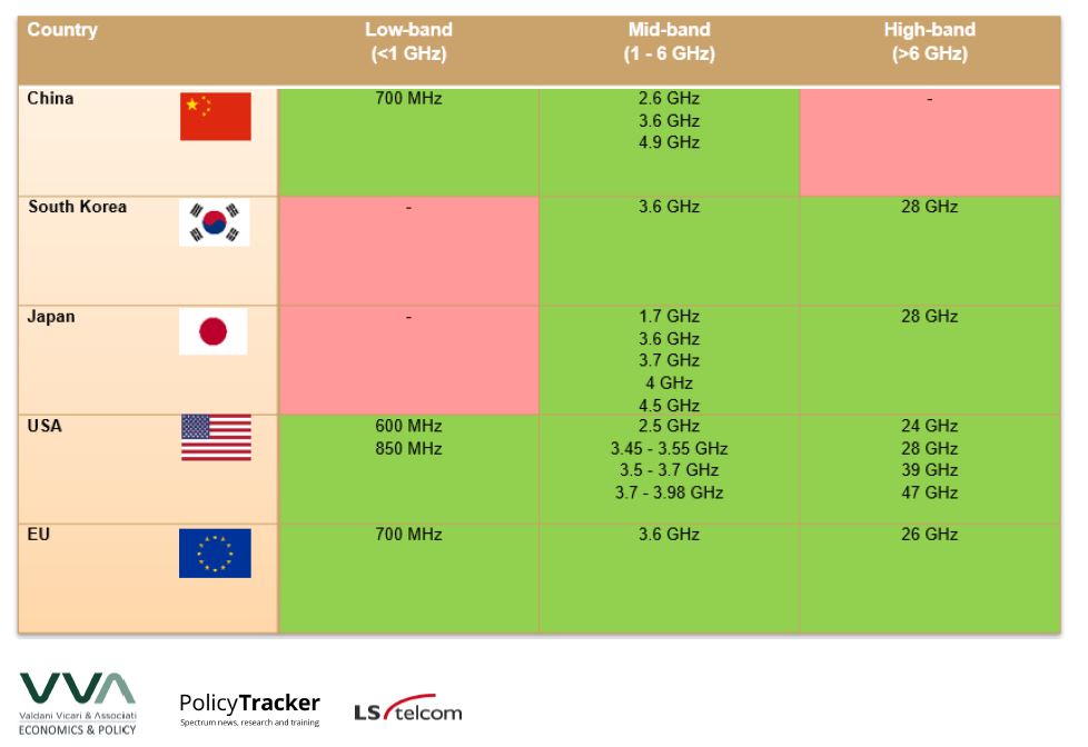
5G Observatory Quarterly Report 13 - Up to October 2021
Valdani Vicari & Associati (VVA), PolicyTracker, LS telcom AG. European Commission Study on “European 5G Observatory phase III." CNECT/2021/OP/0008: 1-135. 2021.
Excerpts
-
In the richest nations like the USA, Japan, South Korea and China,
commercial 5G services have been in operation for a couple of years, and
5G is now spreading to less developed countries.
- The USA has assigned the most mmWave (millimeter wave) spectrum: four bands in total, compared to one in some of the EU and none in China.
-
It is important to note that most of the figures collected on the number
of 5G base stations are provided by governments, but in some cases such
as the USA and Japan, they are based on market research estimates. It
is possible that some market-based estimates are not entirely up to date
or accurate.
==
Possible health effects on the human brain by various
generations of mobile telecommunication: a review based estimation of 5G
impact
Hiie Hinrikus, Tarmo Koppel, Jaanus Lass, Hans Orru, Priit Roosipuu, Maie Bachmann.
Possible health effects on the human brain by various generations of
mobile telecommunication: a review based estimation of 5G impact.
Int J Radiat Biol. 2022 Jan 7;1-48. doi: 10.1080/09553002.2022.2026516.
Abstract
Purpose: The deployment of new 5G NR technology
has significantly raised public concerns in possible negative effects on
human health by radiofrequency electromagnetic fields (RF EMF). The
current review is aimed to clarify the differences between possible
health effects caused by the various generations of telecommunication
technology, especially discussing and projecting possible health effects
by 5G. The review of experimental studies on the human brain over the
last fifteen years and the discussion on physical mechanisms and factors
determining the dependence of the RF EMF effects on frequency and
signal structure have been performed to discover and explain the
possible distinctions between health effects by different
telecommunication generations.
Conclusions: The human
experimental studies on RF EMF effects on the human brain by 2G, 3G and
4G at frequencies from 450 to 2500 MHz were available for analyses. The
search for publications indicated no human experimental studies by 5G
nor at the RF EMF frequencies higher than 2500 MHz. The results of the
current review demonstrate no consistent relationship between the
character of RF EMF effects and parameters of exposure by different
generations (2G, 3G, 4G) of telecommunication technology. At the RF EMF
frequencies lower than 10 GHz, the impact of 5G NR FR1 should have no
principal differences compared to the previous generations. The radio
frequencies used in 5G are even higher and the penetration depths of the
fields are smaller, therefore the effect is rather lower than at
previous generations. At the RF EMF frequencies higher than 10 GHz, the
mechanism of the effects might differ and the impact of 5G NR FR2
becomes unpredictable. Existing knowledge about the mechanism of RF EMF
effects at millimeter waves lacks sufficient experimental data and
theoretical models for reliable conclusions. The insufficient knowledge
about the possible health effects at millimeter waves and the lack of in
vivo experimental studies on 5G NR underline an urgent need for the
theoretical and experimental investigations of health effects by 5G NR,
especially by 5G NR FR2.
Excerpts
Experimental human in vivo studies at radiofrequency range
0.01-300 GHz published in peer-reviewed journals in the last fifteen
years (2007–2021) were eligible, including all types of
telecommunication signals and pulse-modulated radiofrequency radiation.
Altogether 73 publications were included in the review.
According
to investigated parameters, the studies were divided into four
categories: resting electroencephalography (EEG), sleep EEG and sleep
quality, event related potentials (ERP) and cognition-behavior and brain
metabolism. Statistically significant changes in an investigated
parameter between sham and exposed conditions were considered as an
effect.
Table 2 presents the studies that report the RF EMF effect or no effect at different signal structures and frequencies.
No clear interdependency between the generation of telecommunication
technology and the character of RF EMF effects becomes evident from Table 2.
All categories of the reported statistically significant effects as
well as no effects include exposure from various generations of
telecommunication systems and different RF EMF frequencies.
The rate of studies reporting effect is 78.6% at 450 MHz, 66.7% at
900 MHz, 43.6% at 1800 MHz, and 57.1% at 2450 MHz. The rate of positive
findings is maximal, 78.6%, at 450 MHz band and minimal, 43.8%, at
1800 MHz band. However, along with the possible regular frequency
dependent trend, the decrease could be related to other factors:
differences in signal structures and varying number of experiments at
different frequencies. The difference between results at 450 MHz and
1800 MHz can be partly related to the character of applied exposure: at
450 MHz remarkable part of studies have used meander-like
pulse-modulated, not telecommunication signals like RF EMF exposure.
The rate of studies reporting effect is 33.3% at TETRA, 63.6% at GSM,
46.2% at WCDMA, 80% at LTE and 20% at WiFi signals. These numbers should
be taken with caution due to the small number of studies, especially at
LTE, WiFi and TETRA signals. Some trends can be mentioned: the rate of
studies reporting RF EMF effect is higher than 50% at LTE and GSM
signals, lower than 50% at WCDMA and TETRA signals and minimal at WiFi
signals. This trend is not in accordance with the possible dependence on
the used radiofrequency and needs explanation based on the
characteristic behavior of the used signals.
Conclusions
In
the current review, the experimental investigations on RF EMF effects
on human EEG, ERP, cognition and behavior were analyzed at the exposure
conditions typical for the 2G, 3G and 4G generations of mobile
telecommunication technology at frequencies from 450 to 2500 MHz. The
search for publications indicated no studies on human EEG, ERP,
cognition and behavior by 5G nor at RF EMF frequencies higher than
2500 MHz.
The results of the current review demonstrate no
consistent relationship between the character of RF EMF effects and
parameters of exposure by different generations (2G, 3G, 4G) of mobile
telecommunication technology. The following trends can be mentioned:
Various
generations of telecommunication technology seem to contribute to
similar effects. There is no special frequency nor signal structure
related to a specific effect.
Some
decrease in the rate of studies reporting effects with the increase of
RF EMF frequency can be declared. However, due to the small number of
studies, especially at higher frequencies (≥2 GHz), the results need to
be considered with caution.
The existing knowledge about the mechanisms underlying RF EMF effects allows us to formulate the following conclusions:
The
dielectric polarization, a physical reason behind the RF EMF effects,
decreases with the frequency of RF EMF. The electric permittivity is
relatively stable at frequencies over 0.1 and 10 GHz, but decreases fast
at frequencies higher than 10 GHz. At frequencies higher than 10 GHz,
the effects related to the dielectric polarization become small. The
scarce data about the RF EMF effects at frequencies higher than 10 GHz
provide insufficient knowledge to clarify the possible interaction
mechanisms.
The theory of parametric
excitation could explain the impact of the signal structure. The
presence of the low-frequency components lower than 1000 Hz in the
spectrum of RF EMF exposure (2G-5G) is an important factor to give rise
to the RF EMF effects on the nervous system. The RF EMF effects are most
probably caused by the telecommunication systems with low-frequency
components lower than 100 Hz (4G, 5G FR1, 5G FR2).
Currently,
there are no data about RF EMF effects caused by 5G telecommunication
systems. Combining data of experimental results with existing knowledge
in the mechanisms of RF EMF effects, the conclusions about the possible
5G effects can be derived:
At
the RF EMF frequencies lower than 10 GHz, the impact of 5G NR FR1
should have no principal differences compared to the previous
generations. The frequencies used in 5G are even higher and the
penetration depths of the fields are smaller, therefore the effect is
rather lower than at previous generations.
The
low-frequency components in the 5G NR FR1 RF EMF spectrum are similar
to these of 4G. Therefore, the possible health effects should have the
same level.
At the RF EMF frequencies
higher than 10 GHz, the mechanism of the effects might change and the
impact of 5G NR FR2 becomes unpredictable.
The
possible health effects caused by 5G NR FR2 are not limited to the
impact on skin but can be widened by the excitation of nervous system.
Existing
knowledge about the mechanism of RF EMF effects at millimeter waves
lacks sufficient experimental data and theoretical models for reliable
conclusions.
The insufficient knowledge about the possible health effects at millimeter waves and the lack of in vivo
experimental studies on 5G NR underline an urgent need for the
theoretical and experimental investigations of health effects by 5G NR,
especially by 5G NR FR2.
--
Health Effects of 5G Base Station Exposure: A Systematic Review
Tasneem Sofri, Hasliza A Rahim, Mohamedfareq Abdulmalek, Khatijahhusna Abd Rani, Mohd Hafizi Omar, Mohd Najib Mohd Yasin, Muzammil Jusoh, Ping Jack Soh. Health Effects of 5G Base Station Exposure: A Systematic Review. IEEE Access. Dec 30, 2021. doi: 10.1109/ACCESS.2021.3139385.
Abstract
The Fifth Generation (5G) communication technology will deliver faster data speeds and support numerous new applications such as virtual and augmented reality. The additional need for a larger number of 5G base stations has sparked widespread public concerns about their possible negative health impacts. This review analyzes the latest research on electromagnetic exposure on humans, with particular attention to its effect on cognitive performance, well-being, physiological parameters, and Electroencephalography (EEG). While most of their results indicated no changes in cognitive function, physiological parameters, or overall well-being, the strength of the EEG alpha wave is noticed to vary depending on various aspects of cognitive functions. However, the available studies have not investigated the health effects resulting from exposure from the 5G mobile phone and base station antennas from 700 MHz to 30 GHz on the cognitive performance, well-being subjective symptoms, human physiological parameters, and EEG of adults. There is a need for such research regarding this current emerging technology. Such studies are significant in determining whether 5G technology is indeed safe for humans.
Conclusion
This work presents an analysis of exposure studies conducted using signals from 400 MHz to 1750 MHz (for 4G). From this analysis, the following conclusions are made:
• Most of the studies in literature using 2G/3G/4G showed no effects and no consistency in how exposure to these signals affected the cognitive, physiological parameters, well-being, and EEG of the volunteers.
• Most research on human cognition, physiological parameters, and well-being so far have focused on the impacts of GSM900/GSM1800/UMTS/4G MPs, GSM900/GSM1800/UMTS BSs, DECT, and Wi-Fi exposures.
• There is an absence of studies reporting the effects of 5G (700 MHz, 3.5 GHz, or 28 GHz) BS signals on adults in terms of cognitive performance, well-being, or physiological markers (heart rate, blood pressure, and body temperature).
Figure 9 and 10 illustrated the possible flowchart and schematic diagram to study the effects of 5G BS exposure signals for sub-6 GHz and mmWave bands (of up to 30 GHz) to human subjects. Data from such a study will be useful in explicitly determining the significance signal exposure from 5G BS on human health, considering their much closer proximity to users.
--
Health Council of the Netherlands and evaluation of the fifth
generation, 5G,
for wireless communication and cancer risks
Lennart Hardell. Health Council of the Netherlands and evaluation of the fifth
generation, 5G, for wireless communication and cancer risks. World J
Clin Oncol 2021; 12(6): 393-403 doi: 10.5306/wjco.v12.i6.393.
Abstract
Currently the fifth generation, 5G,
for wireless communication is about to be rolled out worldwide. Many
persons are concerned about potential health risks from radiofrequency
radiation. In September 2017, a letter was sent to the European Union
asking for a moratorium on the deployment until scientific evaluation
has been made on potential health risks (http://www.5Gappeal.eu). This
appeal has had little success. The Health Council of the Netherlands
released on September 2, 2020 their evaluation on 5G and health. It was
largely based on a World Health Organization draft and report by the
Swedish Radiation Safety Authority, both criticized for not being
impartial. The guidelines by the International Commission on
Non-Ionizing Radiation Protection were recommended to be used, although
they have been considered to be insufficient to protect against health
hazards (
http://www.emfscientist.org).
The Health Council Committee recommended not to use the 26 GHz
frequency band until health risks have been studied. For lower
frequencies, the International Commission on Non-Ionizing Radiation
Protection guidelines were recommended. The conclusion that there is no
reason to stop the use of lower frequencies for 5G is not justified by
current evidence on cancer risks as commented in this article. A
moratorium is urgently needed on the implementation of 5G for wireless
communication.
Core
Tip: In this comment, guidelines for radiofrequency radiation are
discussed in relation to a recent evaluation by the Health Council of
the Netherlands. The Committee recommends that for the deployment of 5G
the frequency band 26 GHz should not be used. For lower frequencies, the
International Commission on Non-Ionizing Radiation Protection
guidelines are recommended. However, these guidelines are not based on
an objective evaluation of health risks, which is discussed in this
paper.
Conclusion
In conclusion regarding cancer, current scientific evidence clearly
demonstrates an increased risk for glioma and acoustic neuroma for use
of mobile and/or cordless phones. In this review other tumor types and
health endpoints are not discussed. The increased risk for brain and
head tumors is based on human cancer epidemiology studies and is
supported by similar tumor types found in animal studies. In fact, these
animal studies confirmed the earlier results in case-control studies on
increased tumor risk for use of wireless phones (both mobile and
cordless phones). Mechanistic aspects on carcinogenesis come from
laboratory findings on, e.g., the increase of reactive oxygen species[5]
and DNA damage[4].
The current evaluation by the Health Council of the Netherlands is based
on a WHO draft and SSM report. It also recommends using ICNIRP
guidelines, considered to be insufficient to protect against health
hazards, such as cancer, by the majority of the scientists in this field
(https://www.emfscientist.org). The report does not represent a
thorough, balanced, objective, and up-to-date evaluation of cancer risks
and other hazardous effects from RF radiation. It is also strikingly
contradictory as it concludes that serious health effects such as cancer
and birth defects are “possible.” Yet it has no objection to the
roll-out of 5G and recommends that later studies are performed to study
health outcomes such as cancer and birth defects. Thus, no lessons are
learned from existing observations on increased cancer risks[49].
The conclusion by the Commission that there is no reason to stop the use
of lower frequencies for 5G up to 3.5 GHz because of no “proven adverse
health effects,” merely reflects the biased conclusions by ICNIRP
dominated groups. Thus that conclusion must be dismissed, and new
guidelines for previous and new frequencies must be established
considering the new technology, the different propagation pattern for
5G, and increased RF radiation.
A moratorium is urgently required on the implementation of 5G for
wireless communication[13]. Ultimately, wired solutions are preferred.
--
Related Posts
--
Health Safety Guidelines and 5G Wireless Radiation [Health Matters]
James C. Lin. Health Safety Guidelines and 5G Wireless Radiation [Health Matters]. IEEE Microwave Magazine. 23(1):10-17. Jan. 2022, doi: 10.1109/MMM.2021.3117307.
Abstract
The rollout of 5G cellular communication technology is well underway worldwide. The advocates of 5G mobile technology hail it as a faster and more secure technology than its predecessor, 3G and 4G systems. The major enabling infrastructure uses millimeter-wave (mm-wave) and phased-array technology to achieve line-of-sight directivity, high data rates, and low latency. A central vulnerability or security threat is that it may allow spying on users. Nevertheless, this is a system architecture and technology or regulatory issue but not a biological effect or health safety matter.
My note:
James C. Lin, Professor Emeritus in the Department of Electrical and Computer Engineering at the University of Illinois Chicago.
Dr. Lin
is one of the most renowned scientists who has studied the biological
interactions of wireless radiation. He is a fellow of the
American Association for the Advancement of Science and the
Institute of Electrical and Electronics Engineers (IEEE). Since 2006 he has
been the Editor-in-Chief of the
Bioelectromagnetics journal published on behalf of the
Bioelectromagnetics Society (BEMS), an international organization of biological and physical scientists,
physicians and engineers. In a
prior article, Dr.
Lin, an
ICNIRP Commission member from 2004-2016, accused the organization of
groupthink: "The simultaneous penchant to dismiss and criticize positive results and
the fondness for and eager acceptance of negative findings are palpable
and concerning."
Like several previous articles that Dr. JC Lin wrote for IEEE Microwave Magazine, the abstract is biased toward risk minimization so read the paper or the following excerpts.
Excerpts
Low-band 5G starts at roughly 400 MHz and uses existing or previous 3G or 4G frequencies or newly opened frequencies to operate; the latter, for example, may overlap with the existing 4G band. The 5G rollout began with midband, which includes popular frequencies between 3 and 4 GHz. However, primary 5G technological advances are associated with high-band 5G, which promises performance bandwidth as high as 20 GHz, and multiple-input, multiple-output strategies using 64–256 antennas at short distances and offering performances up to 10 times better than the current 4G networks."
"For health safety matters, it is not apparent whether the biological responses to high-band 5G radiations would be akin to earlier generations or low-band 5G radiations, given the distinctive characteristics of mm-wave and its interaction with the complex structure and composition of pertinent, superficial biological cells and tissues such as the cornea of the eye and nerve-rich human skin, the large, protective organ of the body."
"The two most widely promulgated RF health safety guidelines or standards have recently published revisions of their respective 1998 and 2005 versions [1], [2]. The updated International Commission on Nonionizing Radiation Protection guidelines and IEEE standards appear to cater to industry wishes; they are strongly linked to thermal effects associated with measurable temperature elevations. Also, the updates seem to have been synchronized to accommodate the 5G rollout."
"To date, there has not been a single reported epidemiological study that investigated mm-waves and their potential health effects.
Thus, although there are roughly 100 published laboratory investigations of all types, and the reported biological responses are inconsistent in their association between biological effects and mm-wave exposure. Indeed, the types of reported laboratory investigations are small, limited, and diverse, considering the wide, 5G, mm-wave frequency domain. The jury on biological effects or health impacts is still out on 5G mm-waves. Moreover, there is a lack of ongoing, controlled laboratory investigations...."
"If the entities responsible for safety recommendations believe what appears to be their position concerning experimental results from rats from the NIEHS/NTP that a whole-body temperature rise of 1 °C is carcinogenic, then the safety factors of 50 adopted for the public or 10 for workers would be marginal for their stated purpose and practically meaningless from the perspective of “safety” protection (more so above 6 GHz)."
"As shown in Table 1, for mm-waves, the referenced local-tissue-temperature rise in the head, torso, and limbs of humans is 5 °C. This level of temperature rise would bring the tissue temperature from a normal value of 37 °C to a hyperthermic 42 °C. A 42 °C tissue temperature is known to be cytotoxic, with exponential cell-killing capacities. It is used as the basis for clinical cancer therapy in hyperthermia treatment for cancer protocols [14]–[16]. The recently updated safety recommendations provide a reduction factor of 10 for the public’s safety and a reduction factor of two in the case of workers. In this situation, the efficacy of these updated safety recommendations is borderline, and the updated recommendations are meaningless from the perspective of safety protection.
In summary, the safety recommendation updates were based primarily on limiting the tissue-heating potentials of RF radiation to elevate body temperatures. There are significant anomalies in the recently updated safety recommendations. Moreover, aside from the aforementioned anomalies, the existing scientific data are too limited—especially at mm-wavelengths—to make a reliable assessment or conclusion with any certainty. Some of the updated safety recommendations are marginal, questionable, and lack scientific justification from the perspective of safety protection."
--
5G Cellular Standards. Total Radiobiological Assessment of the Danger of Planetary Electromagnetic Radiation Exposure to the Population (in Russian)
Y.G. Grigoriev, A.S. Samoylov. 5G-cellular standards. Total radiobiological assessment of the danger of planetary electromagnetic radiation exposure to the population. G384; М.: SRC — FMBC, Moscow, 2020.
Abstract
The book discusses the implementation of the 5G-standard in the cellular communication system. 5G-technology works with millimeter waves (MMW) with simultaneous distribution of the IoT (Internet of Things) program — Internet connection between «things», both for home use, and other objects, for example, in transport, in production. MMW are easily shielded. Given this, only the skin and sclera of the eyes will be affected.
A new radiobiological approach to hazard assessment of the 5G-standard is presented. The significance of radiobiological criteria and the degree of risk are considered, taking into account the appearance of new critical organs and the load on existing critical organs and systems during lifetime exposure to EMF in the population. This point of view of the authors is used to assess the total radiobiological danger of planetary electromagnetic radiation exposure to the population.
Ways of possible reduction of the electromagnetic load on the population are suggested.
Relevance of the book by L.A. Ilyin, RAS academician
Both in Russia and in countries abroad, there have been active discussions over the past few years about a promising proposal for optimizing cellular communications — the planetary introduction of a new 5G-standard that will guarantee fast transmission of a large amount of data. For this purpose, millimeter waves (MMW) will be used.
The techno-economic advantage of this offer is obvious and widely reported by the media in many countries. However, the degree of danger of this type of electromagnetic radiation to public health and the environment remains unclear.
Unfortunately, the appeals of scientists and medical professionals to the UN and the European Union about the need for preliminary medical and biological research before implementing the 5G-standard remain beyond real implementation. A number of countries refuse to place the 5G-standard on their territory.
Book by Yu.G. Grigoriev and A.S. Samoylov "5G-CELLULAR STANDARD. TOTAL RADIOBIOLOGICAL ASSESSMENT OF THE DANGER OF PLANETARY ELECTROMAGNETIC RADIATION EXPOSURE TO THE POPULATION" considers the implementation of the 5G-standard in the cellular communication system. Unlike existing wireless technologies 2G, 3G and 4G, which use electromagnetic fields of the radio frequency range, the 5G-standard works with millimeter waves with simultaneous distribution of the IoT (Internet of Things) program — Internet communication between "objects", both for home use and other objects, for example, in transport and in production.
For stable delivery of MMW to the entire territory of our planet, Earth satellites are used. It is planned to launch 4,425 satellites for the implementation of the universal Internet access program, but there are already 800 satellites in space under this program. It should be noted that there are currently several thousand satellites in orbit, which is of great concern to astronomers and the security service of manned space flights in Russia.
In fact, the entire population will be trapped for life in the electromagnetic grid of millimeter waves and no one will be able to avoid their impact.
MMW are easily shielded. Naturally, to cover a certain area with a millimeter cell, you will need to increase the number of base stations (BS). For example, with a cell radius of only 20 meters, you will need about 800 base stations per square kilometer and located 3-5 meters from the consumer. This is in sharp contrast, for example, with 3G and 4G-standards, which use large cells and have ranges from 2 to 15 km or more.
Given that MMW is absorbed in biotissues at a depth of up to 2 mm, only the skin and sclera of the eyes will be affected by them. Therefore, the authors rightly believe that when assessing the risk of MMV, it is necessary to take into account the appearance of new critical organs — the skin and eyes. The skin is a very complex biostructure, has a large number of receptors and is actually a "bio-relay" between the external environment and the functional state of the body.
Naturally, the introduction of 5G-technology raises new questions. First, the technical part of providing this type of communication. A significantly larger number of micro-antenna base station antennas per unit area with satellite support is needed. Second, there is a lack of a consistent methodology for hygienic rationing. Third, there are only assumptions about possible biological effects in the lifetime impact of MMW on populations and ecosystems. There are no data on possible bioeffects with constant exposure to MMW on the skin and sclera of the eyes. Targeted research is still not carried out both in Russia and abroad.
There are different perspectives on the assessment of the dangers of this new technology. The International Commission on Non-ionizing Radiation Protection (ICNIRP) and the Federal Communications Commission (FCC) assess the hazard only by adding the absorbed dose to existing standards. This is a small addition, and therefore the existing FCC and ICNIRP standards, approved in 1996, are not being revised. International standards, despite criticism from the scientific community and the European Union, have remained unchanged for more than 20 years.
The authors of the reviewed book consider this approach erroneous, because in this case, the radiation load on new critical organs — the skin and eyes--is not taken into account. They considered the significance of radiobiological criteria and the degree of risk, taking into account the emergence of new critical organs and the load on existing critical organs and systems, taking into account the lifetime exposure of the population to EMF. From this point of view, the book presents an assessment of the total radiobiological danger of planetary electromagnetic radiation exposure to the population.
The book offers new ways to reduce the electromagnetic load, taking into account 5G on the population. It is necessary to explain to the population that EMF is considered harmful and their safety is regulated by certain hygiene standards.
Exposure to EMF that exceeds these standards may negatively affect the health of the mobile user. In this regard, the population should strictly follow the existing hygiene recommendations. However, most people perceive gadgets simply as an element of convenient everyday communication without time limits, as a toy for children, for entertainment, using cellular communication without the need. The population should understand that by violating hygiene recommendations, they are putting themselves at a certain risk. This danger must be persistently explained and, above all, through the media. It is recommended to introduce such a concept as "The conscious risk". This is the first generalization on the problem of the danger of 5G-technologies, both in Russia and abroad.
My comments: A considerable amount of research suggests that exposure to millimeter waves can affect many organs of the body, not just the skin and the eyes.
--
Electromagnetic fields, 5G and health: what about the precautionary principle?
John William Frank.
Electromagnetic fields, 5G and health: what about the precautionary principle? J Epidemiol Community Health. Published Online First: 19 January 2021. doi: 10.1136/jech-2019-213595.
AbstractNew
fifth generation (5G) telecommunications systems, now being rolled out
globally, have become the subject of a fierce controversy. Some health
protection agencies and their scientific advisory committees have
concluded that there is no conclusive scientific evidence of harm.
Several recent reviews by independent scientists, however, suggest that
there is significant uncertainty on this question, with rapidly emerging
evidence of potentially harmful biological effects from radio frequency
electromagnetic field (RF-EMF) exposures, at the levels 5G roll-out
will entail. This essay identifies four relevant sources of scientific
uncertainty and concern: (1) lack of clarity about precisely what
technology is included in 5G; (2) a rapidly accumulating body of
laboratory studies documenting disruptive in vitro and in vivo effects
of RF-EMFs—but one with many gaps in it; (3) an almost total lack (as
yet) of high-quality epidemiological studies of adverse human health
effects from 5G EMF exposure specifically, but rapidly emerging
epidemiological evidence of such effects from past generations of RF-EMF
exposure; (4) persistent allegations that some national
telecommunications regulatory authorities do not base their RF-EMF
safety policies on the latest science, related to unmanaged conflicts of
interest. The author, an experienced epidemiologist, concludes that one
cannot dismiss the growing health concerns about RF-EMFs, especially in
an era when higher population levels of exposure are occurring widely,
due to the spatially dense transmitters which 5G systems require. Based
on the precautionary principle, the author echoes the calls of others
for a moratorium on the further roll-out of 5G systems globally, pending
more conclusive research on their safety.
Conclusions and recommendation
In
assessing causal evidence in environmental epidemiology, Bradford Hill
himself pointed out that ‘the whole picture matters;’ he argued against
prioritising any subset of his famous nine criteria for causation. One’s
overall assessment of the likelihood that an exposure causes a health
condition should take into account a wide variety of evidence, including
‘biological plausibility’. After reviewing the evidence cited above, the writer, an experienced
physician-epidemiologist, is convinced that RF-EMFs may well have
serious human health effects. While there is also increasing scientific
evidence for RF-EMF effects of ecological concern in other species, both plant and animal, these have not been reviewed here, for reasons
of space and the author’s disciplinary limitations. In addition, there
is convincing evidence, cited above, that several nations’ regulatory
apparatus, for telecommunications innovations such as the 5G roll-out,
is not fit for purpose. Indeed, significant elements in that apparatus
appear to have been captured by vested interests. Every society’s public
health—and especially the health of those most likely to be susceptible
to the hazard in question (in the case of EMFs, children and pregnant
women)—needs to be protected by evidence-based regulations, free from
significant bias.
Finally, this commentary would be
remiss if it did not mention a widely circulating conspiracy theory,
suggesting that 5G and related EMF exposures somehow contributed to the
creation or spread of the current COVID-19 pandemic. There are
knowledgeable commentators’ reports on the web debunking this theory, and no respectable scientist or publication has backed it. Indeed, combatting it is widely viewed by the scientific community as critical to dealing with the pandemic, as conspiracy theorists holding this view have already carried out violent attacks on mobile phone transmission facilities and other symbolic targets, distracting the public and authorities at a time when pandemic control actions are paramount. 42 This writer completely supports that view of the broader scientific community: the theory that 5G and related EMFs have contributed to the pandemic is baseless.
It follows that, for the current 5G roll-out, there is a sound basis for invoking ‘the precautionary principle’. This is the environmental and occupational health principle by which significant doubt about the safety of a new and potentially widespread human exposure should be a reason to call a moratorium on that exposure, pending adequate scientific investigation of its suspected adverse health effects. In short, one should ‘err on the side of caution’. In the case of 5G transmission systems, there is no compelling public health or safety rationale for their rapid deployment. The main gains being promised are either economic (for some parties only, not necessarily with widely distributed financial benefits across the population) or related to increased consumer convenience. Until we know more about what we are getting into, from a health and ecological point of view, those putative gains need to wait.
Open access paper: https://jech.bmj.com/content/early/2021/01/04/jech-2019-213595 or https://jech.bmj.com/content/jech/early/2021/01/04/jech-2019-213595.full.pdf
--
Chemical polarization effects of electromagnetic field radiation from the novel 5G network deployment at ultra high frequency
Ugochukwu O. Matthew, Jazuli S. Kazaure. Chemical polarization effects of electromagnetic field radiation from the novel 5G network deployment at ultra high frequency. Health Technology (Berl). 2021 Jan 27: 1-13. doi: 10.1007/s12553-020-00501-x.
Abstract
The wide-spectrum of
non-ionizing, non-visible radiation emitted from the novel 5G network
deployment was investigated and found liable to produce effects capable
of heating up and altering human body nomenclature. The Ultra-high
frequency magnetic fields, induced circulation of currents in the
surrounding human body when potentially exposed. The quantum of these
electromagnetic charges is influenced by the magnitude of the external
magnetic field. The Magnetic fields warming is the major organic
consequence of the electromagnetic fields radiofrequency radiation
emitted from 5G network installation especially at a very high
frequencies. From the current research, the levels of electromagnetic
fields to which individuals are naturally unmasked under 4G network and
5G network technology in SCENARIO1, SCENARIO 2 and SCENARIO 3 are very
negligible to alter human body dipolar chemistry. On the several
findings of the research, deploying 5G network technology under the
ultra-high frequency above 20 GHz will produce effect that will heat up
the human body tissues due to electromagnetic field inducement since
human body is dipolar in nature. The research established that while the
current digital society will continue investment into 5G network
technology, caution must be applied not to deploy 5G network under
ultra-high frequency above 20 GHz due to its adverse health effects.
ConclusionsFrom
the knowledge and principle of electromagnetism, human beings are
constituted of substantial amount of oriented cells with diverse
electromagnetic field attributes. The Biological attributes of the human
tissue under diverse electromagnetic radiative emission are studied and
that had provided the basis upon which the current research on the
effects of electromagnetic fields on the human body. The heating
consequences of the radio electromagnetic waves from 5G network
technology deployment had formed the fundamental basis for current
research. On the several findings of the research, deploying 5G network
technology under the ultra-high baseband above 20 GHz will produce
effects such as heating up of the body tissues due to electromagnetic
field inducement on the account that human body is dipolar in nature.
The effects will extend to produce dielectric polarization, ionic
polarization, interfacial polarization and orientational polarization.
This is generally on the account that variations on dielectric
properties of biological tissues with the frequency of the
electromagnetic field inducement are very dissimilar. While it is very
imperative to determine the frequency distribution in deploying the
novel 5G network to avoid adverse dielectric dispersion that may flow
into the human body.
Open access paper: https://www.springerprofessional.de/en/chemical-polarization-effects-of-electromagnetic-field-radiation/18805704
--
Abstract
The
fifth-generation wireless (5G) has already started showing its
capability to achieve extremely fast data transfer, which makes itself
considered to be a promising mobile technology. However, concerns have
been raised on adverse health impacts that human users can experience in
a 5G system by being exposed to electromagnetic fields (EMFs). This
article investigates the human EMF exposure in a 5G system and compares
them with those measured in the previous-generation cellular systems. It
suggests a minimum separation distance between a transmitter and a
human user for keeping the EMF exposure below the safety regulation
level, which provides consumers with a general understanding on the safe
use of 5G communications.
Excerpts
"First, we discuss the human EMF exposure in the downlink as
well as the uplink. Most of the prior work studies the uplink only,
while hardly paying attention to EMF emissions generated by BSs
[base stations or cell towers]
in a 5G
network. Recall the aforementioned changes that the 5G adopts: 1)
operation at higher carrier frequencies; 2) reduction of cell size
(which leads to increase in number of BSs; and 3) concentration of
higher EMF energy into an antenna beam. They all imply that in 5G,
unlike the previous-generation wireless systems, the downlink can also
be a threat to human health as well as the uplink.
Second, we suggest that both SAR [Specific Absorption Rate] and PD
[power density] should be used to display human EMF exposure for a wireless system. The
reason is that SAR captures an amount of EMF energy that is actually
“absorbed” into human tissues, whereas PD is an efficient metric only to
present the EMF energy being introduced to a human user.
Third, we present an explicit comparison of human EMF exposure in 5G to those in the currently deployed wireless standards....
Fourth, we consider the maximum possible exposure that a human user can experience...."
"... in a 5G network, a consumer is likely to be exposed to high EMF energy
more consistently. Nevertheless, it is easier to apply a “compliance
distance” [17]
in a downlink than in an uplink. Thus, this article suggests 1) an
overhaul of the compliance distances defined in different standards and
2) the consumers’ discretion on being close to a BS...."
"... the
fact that a high-frequency EMF cannot penetrate deep into human skin
does not mean that it is not dangerous. Specifically, although the
penetration is limited only at the skin surface, the SAR (illustrated as
a heat map in Figure 4) can be higher within the concentrated area, which can cause subsequent health problems such as skin heating."
Downlink vs. Uplink
"Figure 3(c) and (d) compare PD and SAR in uplink to the ICNIRP guidelines set at 10 W/m2 and 2W/kg, respectively. PD and SAR are remarkably higher in uplink than those in downlink, shown via a comparison of the results for uplink to those for downlink shown in Figure 3(a) and (b). It is attributed to smaller separation distance between a transmitter and a human body. Imagine one talking on a voice call; it is a “direct” physical contact of the phone and the head!
Also, it is significant to notice that no regulation exists at 28 GHz where this article investigates for 5G. As such, we refer to the ICNIRP's guideline that is set to be 2 W/kg by ICNIRP[11] at a frequency “below 10 GHz.” In Figure 3(d), it provides a ““inferred” understanding on SAR in an uplink. The zoom-in look shown in Figure 3(d) suggests that in 5G, use of a handheld device within the distance of 8 cm causes an EMF absorption exceeding 2 W/kg, which would have been prohibited if the carrier frequency was lower than 10 GHz. This implies the gravity of human EMF exposure in an uplink of 5G."Conclusion
"This article has discussed human EMF exposure in 5G operating at
28 GHz, while most of the prior work focuses only on the technological
benefits that the technology brings. Considering the significance of
wireless technologies in our daily life, the potential danger of using
them should also be emphasized for sustainable advancement of the
technologies. In this article, the first case study has demonstrated how
much EMF exposure is caused in a 5G system compared to 4G and 3.9G.
Then, the latter case study has suggested an adequate separation
distance from a transmitter, in order to keep a human user from being
exposed to EMF below a regulatory guideline. This article is expected to
ignite continued interest in overarching research on the design of
future wireless systems that achieve high performance while keeping
consumer safety guaranteed.
However, considering the gravity of this issue, we suggest several directions to be achieved in our future research.
Human EMF exposure mitigation strategy:
We are particularly interested in exploiting the technical features in
future wireless systems—i.e., a larger number of BSs within a unit area.
Such a paradigm change will enable a holistic, network-based approach
to mitigate the EMF exposure as an optimization problem with a set of
constraints representing the PD, SAR, and skin-temperature elevation.
Further studies regarding exact human health impacts caused by EMF exposure:
The particular focus will be put on 1) skin dielectric effect with
respect to frequency and 2) the effect of radiation when the body is
covered with clothing or garment materials."
--
Modelling of Total Exposure in Hypothetical 5G Mobile Networks
for Varied Topologies and User Scenarios
Sven Kuehn, Serge Pfeifer, Beyhan Kochali, Niels Kuster.
Modelling of Total Exposure in Hypothetical 5G Mobile Networks for Varied Topologies and User Scenarios. Final Report of Project CRR-816. A report on behalf of the Swiss Federal Office for the Environment (FOEN). Zurich, IT'IS Foundation. 24 June 2019.
Executive Summary
In January 2019, the Swiss Federal Office for the Environment (FOEN) mandated the IT’IS Foundation to evaluate the total human exposure in hypothetical 5G mobile networks for varied topologies and user scenarios to identify factors that would minimize the total exposure of the population. In this study, total exposure is defined as the combined exposure from network base stations, the user’s own device, as well as bystanders’ mobile devices.
The influence of various factors on total exposure in mobile communication networks (as defined above) was modeled and analyzed with the help of the Monte Carlo simulation technique. Total exposure is described as the local peak specific absorption rate (SAR) spatially averaged over any 10 g of tissue mass (psaSAR10g) averaged over a period of 6 minutes. The unit psaSAR10g was chosen because it defines the governing basic restriction for wireless exposure as the whole-body average SAR limits (wbaSAR) are intrinsically met if the limits of local exposure are satisfied. The averaging duration of 6 minutes constitutes the internationally accepted averaging time to prevent thermal hazards at frequencies below 6 GHz as instant values have little justification. However, it should be noted that some regulators define shorter averaging time periods, e.g., the US Federal Communications Commission (FCC) of 100 s.
In a first step, we analyzed the tissue-specific exposure as a function of frequency. The preliminary dosimetric study showed that exposure of the human brain to the 3.6 GHz band, that has been recently added to the Swiss mobile communication frequencies, is reduced by a factor of >6 for the tissue averaged SAR when compared to mobile network operation at <1 GHz. This reduction is due to the smaller penetration depth at higher frequencies. This conclusion, however, does not apply to exposed tissues close to the surface or skin (eyes, testicles, etc.) when the peak SAR in this tissue is evaluated. The peak SAR in the grey matter remains in approximately the same order of magnitude (�3 dB) over all frequencies but the area of high exposure is reduced at 3.6 GHz.
In a second step, we used data measured in 4G systems and analyzed the latest mobile network standards to extrapolate the exposures for various 5G network scenarios. These measured data were also used to extrapolate the exposure to the future development of data usage in 5G networks.
Specifically, we analyzed the effect on the total exposure of (i) the network topology by varying the cell size and amount of indoor coverage in the network, as well as the usage of (ii) an individual’s own device, and (iii) devices of close bystanders.
The results – based on simulations of more than 200 different exposure scenarios – reveal that, for all user types, except for non-users (including passive mobile phone users and users dominantly using downlink data traffic, e.g., video streaming), total exposure is dominated by the person’s own mobile device. Compared to non-users, the exposure is increased (i) for light users (with 100 MByte uplink data per day) by 6 – 10 dB (or a factor of 4 to 10), (ii) for moderate users (with 1 GByte uplink data per day) by 13 – 25 dB (or a factor of 20 to >300), and (iii) for heavy users by 15 – 40 dB (or a factor of 30 to >10000). Further, the results show that peak exposure of non-users is not defined by exposure to base stations but by exposure to mobile devices of close bystanders in urban areas resulting in 6 dB (or a factor of 4) higher exposure than from a nearby base station antenna.
While a reduction of the mobile cell size leads to a reduction in total exposure by a factor of 2 to10 for people actively using their mobile devices, this might also lead to a small increase by a factor of 1.6 in total exposure of non-users due the generally increased incident signal levels from the surrounding base stations.
Similarly, the exposure of active users can be reduced by a factor of 4 to 600 by increasing the indoor network coverage. Yet, in line with the results for the mobile cell sizes, increased indoor coverage will also lead to increased exposure of non-users by a factor of 2 to 10. This increase, however, starts at a level 1000 times lower than the typical total exposure of active users.
The results of this study show that the personal mobile device is the dominant exposure source for active mobile network users. Besides a person’s own usage behavior, total exposure is also closely linked to the network infrastructure. Generally speaking, a network with a lower path loss, i.e., smaller cells and additional indoor coverage, helps to reduce total exposure. The exposure per transmitted bit is reduced by a factor of <3 by the increased spectral efficiency of the 5G technology, and the reduced penetration depth associated with the new bands at 3.5 – 3.8 GHz.
The results presented above are limited due to the network data that has been used and the definition of total exposure as stated in this report. Furthermore, it only considers time-averaged (6 min) and not instant exposures. This study does not consider (i) the effect of upcoming massive MIMO systems in 5G networks, (ii) alternative data transmission links, for instance the use of Wireless Local Area Network (WLAN), and (iii) millimeter wave frequencies in 5G mobile networks.
Conclusions
The results of this study show that the absorption of energy by the human brain, resulting from exposure to the 3.6 GHz band newly added to the Swiss mobile communication frequencies, is reduced by a factor >6 for the tissue averaged SAR when compared to mobile networks operating at <1 GHz, and by a factor of >2 when compared to the frequency bands at 1.8 – 2GHz. For deep brain regions, the reduction is much larger.
The reduced exposure for these regions is due to lower penetration depths at higher frequencies. Close to the surface (eyes, testicles, etc.) the exposure can be higher. At the most exposed surface of the grey matter, the values remain approximately �3 dB over all frequencies whereas the area of high exposure is reduced.
More than 200 Monte Carlo simulated exposure scenarios have been analyzed to evaluate total human exposure in 5G Networks for different topologies and user scenarios. The results show that for all users (except non-users), the total exposure is dominated by a person’s own mobile device. Compared to a non-user, the exposure is increased for a light user (with 100 MByte uplink data per day) by 6 – 10 dB (or by a factor 4 to 10), for a moderate user (with 1 GByte uplink data per day) by 13 – 25 dB (or by a factor of 20 to >300), and for a heavy user by 25 – 40 dB (or a factor of 300 to >10000). The peak exposure of non-users is further not defined by exposure to surrounding base stations but by mobile devices of close bystanders in urban areas, resulting in 6 dB (or a factor of 4) higher exposure than from a nearby base station antenna.
Reducing the diameter of the mobile cell leads to a decreased overall exposure by a factor of 2 to 10 for people who actively use their mobile devices. At the same time, the reduction in cell size might lead to a small increase by a factor <2 in exposure for non-users. The exposure of active users can be reduced by factors ranging from 4 to 600 by increasing indoor network coverage which, in turn, will be linked to increased exposure of non-users by a factor of 2 to 10. However, such an increase is by a factor 1000 lower than the typical exposure of active users. The results of this study are limited due to the network data that has been used and the definition of total exposure as stated earlier in this report. This study does not consider (i) the effect of upcoming massive MIMO and multi-user MIMO systems in 5G networks, (ii) alternative data transmission links – for instance the use of Wireless Local Area Network (WLAN) and (iii) millimeter wave frequencies in 5G mobile networks.
In summary, the results of this study show that the user’s own mobile device is the dominant source of exposure for the population of active mobile network users. Besides personal usage patterns, totl exposure is also closely linked to the network infrastructure. Generally speaking, a network that decreases the path loss by means of smaller cells and additional indoor coverage will help to reduce the total exposure of the population.
--
Oct 14, 2020
5G Research
from the EMF-Portal Archive
As of June 1, 2020, the EMF-Portal archive listed 133 papers and letters to the editor published in professional journals and
presentations at professional conferences that focus on 5G research. Although most
discuss technical or dosimetric issues (n = 92), 41 citations address other
issues including potential biologic or health effects.
Currently, no peer-reviewed, empirical studies of the biologic
or health effects from actual exposure to 5G radiation have been published. Hence,
those who claim that 5G is safe because it complies with radiofrequency
exposure guidelines are engaging in sophistry.
These guidelines were designed to protect the
population from short-term heating (or thermal) risks. However, numerous peer-reviewed
studies have found adverse biologic and health effects from exposure to low-intensity
or non-thermal levels of electromagnetic fields (EMF). Hence, more than 240 EMF
scientists who have signed the International EMF Scientist Appeal have recommended that “guidelines and regulatory standards be strengthened”:
“Numerous recent scientific publications have shown
that EMF affects living organisms at levels well below most international and
national guidelines….
The
various agencies setting safety standards have failed to impose sufficient
guidelines to protect the general public, particularly children who are more
vulnerable to the effects of EMF.”
--
5G Wireless Deployment and Health Risks: Time for a Medical Discussion
Priyanka
Bandara, Tracy Chandler, Robin Kelly, Julie McCredden, Murray May,
Steve Weller, Don Maisch, Susan Pockett, Victor Leach, Richard Cullen,
Damian Wojcik.
5G Wireless Deployment and Health Risks: Time for a Medical Discussion
in Australia and New Zealand. ACNEM Journal. 39(1). July 2020.
No abstract.
Excerpts
"There is an urgent need for clinicians and
medical scientists in the Australia-New Zealand region to engage in an
objective discussion around the potential health impacts of the fifth
generation (5G) wireless technology currently being deployed. The
statements of assurance by the industry and government parties that
dominate the media in our region are at odds with the warnings of
hundreds of scientists actively engaged in research on biological/health
effects of anthropogenic electromagnetic radiation/fields (EMR/EMF).
(1) There have been worldwide public protests as well as appeals by
professionals and the general public (2) that have compelled many cities
in Europe to declare moratoria on 5G deployment and to begin
investigations. In contrast, there is no medically-oriented
professional discussion on this public health topic in Australia and New
Zealand, where 5G deployment is being expedited. 5G is untested for
safety on humans and other species and the limited existing evidence
raises major concerns that need to be addressed. The vast body of
research literature on biological/health effects of ‘wireless
radiation’ (radiofrequency EMR) (3,4) indicates a range of
health-related issues associated with different types of wireless
technologies (1G-4G, WiFi, Bluetooth, Radar, radio/TV transmission,
scanning and surveillance systems). These are used in a wide range of
personal devices in common use (mobile/cordless phones, computers, baby
monitors, games consoles etc) without users being aware of the health
risks. Furthermore, serious safety concerns arise from the extra
complexity of 5G as follows:
•
5G carrier waves use a much broader part of the microwave spectrum
including waves with wavelengths in the millimetre range (hence called
‘millimetre waves’) which will be used in the second phase of 5G).
Until now, millimetre waves have had limited applications such as radar,
point-to-point communications links and non-lethal military weapons.
(5)
• Extremely complex modulation patterns involving numerous frequencies form novel exposures.
• Beam formation characteristics can produce hotspots of high unknown intensities.
•
A vast number of antenna arrays will add millions of microwave
transmitters globally in addition to the existing RF transmitters
thereby greatly increasing human exposure. This includes 5G small cell
antennas to be erected every 200-250 metres on street fixtures, such as
power poles and bus shelters, many of which will be only metres from
homes with the homeowners having absolutely no say in where the antennas
will be located.
This massive leap in human
exposure to RF-EMR from 5G is occurring in a setting where the existing
scientific evidence overwhelmingly indicates biological interference,
(3,4) therefore suggesting the need to urgently reduce exposure...."
"As
for the new 5G technology, it is concerning that leading experts in the
technical field (6) have reported the possibility of damaging thermal
spikes under the current exposure guidelines (from beam forming 5G
millimetre waves that transfer data with short bursts of high energy)
and some animals and children may be at an increased risk due to smaller
body size. Even working within the entirely thermally-based current
regulatory process, they pointed out 5G millimetre waves “may lead to
permanent tissue damage after even short exposures, highlighting the
importance of revisiting existing exposure guidelines”. (6) Microwave
experts from the US Air Force have reported on ‘Brillouin Precursors’
created by sharp transients at the leading and trailing edges of pulses
of mm waves, when beam forming fast millimetre waves create moving
charges in the body which penetrate deeper than explained in the
conventional models, and have the potential to cause tissue damage. (7)
In fact, concerns about moving charges affecting deep tissue are
associated with other forms of pulsed RF radiation currently used for
wireless communications. This may be one factor explaining why the
pulsed radiation used in wireless communication technologies is more
biologically active than continuous RF radiation. (8) Such effects of
high energy 5G mm waves could have potentially devastating consequences
for species with small body size and also creatures that have innate
sensitivity to EMF, which include birds and bees that use nature’s EMFs
for navigation. (9) Unfortunately, non-thermal effects and chronic
exposure effects are not addressed in the current guidelines. (10)"
"Our
investigation into the scientific literature has found RF-EMR to be a
potent inducer of oxidative stress even at so-called “low-intensity”
exposures (which are in fact billions of times higher than in nature
(26)) such as those from commonly used wireless devices. An analysis
(22) of 242 publications (experimental studies) which had investigated
endpoints related to oxidative stress - biomarkers of oxidative damage
such as 8-oxo-2'-deoxyguanosine (indicating oxidative DNA damage) and/or
altered antioxidant levels - revealed that 216 studies (89%) had
reported such findings (Fig. 1). This evidence base on RF-associated
oxidative stress from 26 countries (only one study from Australia and
none from New Zealand) is relatively new and mostly post 2010, i.e.
after the WHO’s International Agency for Research on Cancer (IARC)
classified RF-EMR as a Group 2B possible carcinogen. Moreover, 180
studies out of the 242 (74.7%) were in vivo studies (including several
human studies) which presents strong evidence.
"Proponents
of 5G often dismiss concerns about health risks claiming that 5G
microwaves will minimally penetrate the skin and therefore any effects
are limited to minor skin heating (and they acknowledge that there is
some uncertainty around heating effects on the eyes). The medical
community understands that skin is the largest organ of the human body
and a key part of the neuro-immune and neuro-endocrine systems. Natural
UVA and UVB (also so-called non-ionizing radiation) that penetrate the
skin less than 5G millimetre waves have profound effects on health and
wellbeing of humans. Therefore, artificial 5G waves must be subjected to
rigorous safety testing."
"Unfortunately, the
questionable conduct of regulatory agencies such as ARPANSA and WHO’s
international EMF Project (43) with conflicts of interest due to funding
links to the wireless industry (44) remains to be investigated. More
open questioning and protests are appearing in Europe and North America
where there is some level of engagement on the part of government bodies
in response to warnings of adverse health effects of anthropogenic
EMF/EMR by expert medical bodies such as EUROPAEM and AAEM (31,32)
(despite industry opposition)."
--
5G Communication Technology and Coronavirus Disease [Health Matters]
James C. Lin. 5G Communication Technology and Coronavirus Disease [Health
Matters]. IEEE Microwave Magazine, 21(9):16-19. Sep 2020.
No abstract.
Excerpts
"The fact is that there is no link between the COVID-19 virus and 5G cell phone technology or 5G base-station communication towers. These are totally different constructs; they are not even close. None of the conspiracy theories that try to link 5G and the coronavirus make any sense scientifically."
"For biological matters, it is not obvious whether the biological responses to high-band 5G radiation will be akin to earlier generations or low-band 5G radiations, given the distinctive characteristics of mm-wave [millimeter wave] and its interaction with the complex structure and composition of pertinent biological tissues."
"It is important to note that the recent NTP and Ramazzini RF exposure studies presented similar findings in terms of heart schwannomas and brain gliomas. Thus, two relatively well-conducted RF exposure studies employing the same strain of rats showed consistent results in significantly increased cancer risks. More recently, an advisory group for the IARC has recommended including reevaluation of the carcinogenicity of human exposure to RF radiation, with high priority, in their monograph series [7]."
"... the 5G frequency domain is divided into low, mid, and high bands. The operating frequencies at low and mid bands can overlap with the current 4G band at 6 GHz or below. Thus, the biological effects of RF radiation at these lower frequency bands are likely to be comparable to 2, 3, or 4G. However, the scenarios of high-band 5G—especially for 24–60 GHz in the mm-wave region for high-capacity, short-range wireless data communications—are relatively recent arrivals and pose considerable challenge to health risk assessment. There is a paucity of data on permittivity and coupling, such as reflection, transmission, and induced energy deposition, in biological tissues in the mm-wave frequency band."
"Induced energy deposition increases with mm-wave frequency. However, at the highest frequencies, the energy deposition in the deeper regions inside the skin is lower because of the reduced penetration depth at these frequencies [11]."
"A recently published review [13] included 45 in vivo studies conducted using laboratory animals and other biological preparations and 53 in vitro studies involving primary cells and cultured cell lines.... This industry-supported review noted that, aside from the wide frequency ranges, the studies were diverse both in subjects and in the end points investigated. Biological effects were observed to occur both in vivo and in vitro for different biological endpoints studied. Indeed, the percentage of positive responses at nonthermal levels in most frequency groups was as high as 70%."
"While many of these investigations with mm-wave exposures reported biological responses, there is inconsistency in the dependence of biological effects and mm-wave intensity used for exposure. Also, the reported in vitro and in vivo laboratory investigations are modest in number and diverse in subject matter, considering the wide 5G/mmwave frequency domain. The jury on the biological effect or health impact is still out on 5G. Moreover, there is a lack of ongoing controlled laboratory investigations. Simply put, the existing scientific data are too limited for any reliable assessment or conclusion with certainty."
--
Setting
Guidelines for Electromagnetic Exposures and Research Needs
Barnes F,
Greenebaum B. Setting Guidelines for Electromagnetic Exposures and Research
Needs. Bioelectromagnetics. 2020 Jul;41(5):392-397. doi: 10.1002/bem.22267.
Abstract
Current limits for exposures to nonionizing electromagnetic fields (EMF) are
set, based on relatively short-term exposures. Long-term exposures to weak EMF
are not addressed in the current guidelines. Nevertheless, a large and growing
amount of evidence indicates that long-term exposure to weak fields can affect
biological systems and might have effects on human health. If they do, the
public health issues could be important because of the very large fraction of
the population worldwide that is exposed. We also discuss research that needs
to be done to clarify questions about the effects of weak fields. In addition
to the current short-term exposure guidelines, we propose an approach to how
weak field exposure guidelines for long-term exposures might be set, in which
the responsibility for limiting exposure is divided between the manufacturer,
system operator, and individual being exposed.
Excerpts
“Both IEEE and
ICNIRP base their analyses on rigorous reviews of the scientific literature and
on established firm evidence of health effects in humans. The present
guidelines are based on acute exposures; to date both IEEE and ICNIRP have not
found sufficient evidence to include health effects of long-term exposures at
lower levels. However, over the last 20 years the evidence has become extremely
strong that weaker EMF over the whole range for frequencies from static through
millimeter waves can modify biological processes. There is now solid
experimental evidence and supporting theory showing that weak fields,
especially but not exclusively at low frequencies, can modify reactive free
radical concentrations and that changes in radical concentration and that of
other signaling molecules, such as hydrogen peroxide and calcium, can modify
biological processes …”
“The evidence
that weak radiofrequency (RF) and low-frequency fields can modify human health
is still less strong, but the experiments supporting both conclusions are too
numerous to be uniformly written off as a group due to poor technique, poor
dosimetry, or lack of blinding in some cases, or other good laboratory
practices. Based on recent studies by the National Toxicology Program (NTP)
[Smith‐Roe et
al., 2020] and the Ramazzini Foundation [Falcioni et al., 2018] as well as
laboratory data, the International Agency for Research on Cancer (IARC) has
declared RF fields as possible human carcinogens [IARC, 2013]. A recent paper
extends the NTP studies by evaluating genotoxicity in animals exposed to fields
at or over the guideline limits and found DNA damage in Comet assays [Smith‐Roe et al., 2020]. Many other papers
indicate similar results, but many negative results are also in the
literature.”
“PROPOSED APPROACH TO SETTING EXPOSURE LIMITS
From these and other lines of solid research, the guidelines for exposure could
be revised. Increased emphasis on long-term exposures may require refining the
concept of dose to more flexibly combine exposure time and field intensity or
energy absorbed. Eventual guidelines might suggest limiting cell phone calls to
X hours per day with exposure levels above Y W/m2, and for Z days per week
exposure should be less than Y W/m2 to allow the body to reset its baseline.”
“What is missing in the current guidelines or regulations are guidelines for
long-term exposure to weak EMF….”
“Guidelines should be set at three levels: the individual user, local company,
and national or international level…. External guidance, in terms of informed
recommendations or at least analysis of various intensities and styles of usage
from some agency such as the Federal Communications Commission (FCC) or NIH,
would be useful.
Limits on the time for operations of base stations and exposures in adjacent
living spaces are not controlled by the user and must be set by competent
authorities, based on scientific evidence. It is likely to be difficult to
specify times when exposures to RF signals are zero or below some limit. What
will be needed is being able to say with some certainty that exposure below a
given level has not been shown to cause changes in body chemistry above some
level. A starting point might be current levels from TV and radio stations that
are large enough to give signal-to-noise ratios around 20 dB (100-fold) with
typical receiving systems. Currently, mean values for the population's exposure
to these systems are estimated to be around 0.1 V/m and peak exposures range up
to 2 V/m, which exceed current exposure limits for a small fraction of the
population. Therefore, one starting point for exposure limits might be an
average of 0.1 V/m, not based on research but on practicality, until further
research results dictate either a lower or higher limit.”
--
Effects of 5G Wireless Communication on Human Health
Karaboytcheva M. Effects of 5G wireless communication on human health. European Parliamentary Research Service (EPRS). Briefing document: PE 646.172. March 2020.
Summary
The fifth generation of telecommunications technologies, 5G, is fundamental to achieving a European gigabit society by 2025.
The aim to cover all urban areas, railways and major roads with uninterrupted fifth generation wireless communication can only be achieved by creating a very dense network of antennas and transmitters. In other words, the number of higher frequency base stations and other devices will increase significantly.
This raises the question as to whether there is a negative impact on human health and environment from higher frequencies and billions of additional connections, which, according to research, will mean constant exposure for the whole population, including children.
Whereas researchers generally consider such radio waves not to constitute a threat to the population, research to date has not addressed the constant exposure that 5G would introduce. Accordingly, a section of the scientific community considers that more research on the potential negative biological effects of electromagnetic fields (EMF) and 5G is needed, notably on the incidence of some serious human diseases. A further consideration is the need to bring together researchers from different disciplines, in particular medicine and physics or engineering, to conduct further research into the effects of 5G.
The EU’s current provisions on exposure to wireless signals, the Council Recommendation on the limitation of exposure of the general public to electromagnetic fields (0 Hz to 300 GHz), is now 20 years old, and thus does not take the specific technical characteristics of 5G into account.
In this Briefing
- Difference between 5G and current technology
- Regulation of electromagnetic fields and 5G exposure
- European Parliament Research on EMF and 5G effects on human health
- Stakeholders' views
- The road ahead for 5G
--
Adverse health effects of 5G mobile networking technology under real-life conditions
Kostoff RN, Heroux P, Aschner M, Tsatsakis A. Adverse health effects of 5G mobile networking technology under real-life conditions. Toxicology Letters. 323(1):35-40. May 2020. https://doi.org/10.1016/j.toxlet.2020.01.020.
Highlights
• Identifies wide-spectrum of adverse health effects of non-ionizing non-visible radiation
• Most laboratory experiments were not designed to identify the more severe adverse effects reflective of real-life conditions
• Many experiments do not include the real-life pulsing and modulation of the carrier signal
• Vast majority of experiments do not account for synergistic adverse effects of other toxic stimuli with wireless radiation
• 5G mobile networking technology will affect not only the skin and eyes, but will have adverse systemic effects as well
Abstract
This article identifies adverse effects of non-ionizing non-visible radiation (hereafter called wireless radiation) reported in the premier biomedical literature. It emphasizes that most of the laboratory experiments conducted to date are not designed to identify the more severe adverse effects reflective of the real-life operating environment in which wireless radiation systems operate. Many experiments do not include pulsing and modulation of the carrier signal. The vast majority do not account for synergistic adverse effects of other toxic stimuli (such as chemical and biological) acting in concert with the wireless radiation. This article also presents evidence that the nascent 5G mobile networking technology will affect not only the skin and eyes, as commonly believed, but will have adverse systemic effects as well.
Appeals that matter or not on a moratorium on the deployment of the fifth generation, 5G, for microwave radiation
Hardell L, Nyberg R. [Comment] Appeals that matter or not on a moratorium on the deployment of the fifth generation, 5G, for microwave radiation. Molecular and Clinical Oncology. Published online January 22, 2020. https://doi.org/10.3892/mco.2020.1984.
Abstract
Radiofrequency (RF) radiation in the frequency range of 30 kHz‑300 GHz is classified as a ‘possible’ human carcinogen, Group 2B, by the International Agency for Research on Cancer (IARC) since 2011. The evidence has since then been strengthened by further research; thus, RF radiation may now be classified as a human carcinogen, Group 1. In spite of this, microwave radiations are expanding with increasing personal and ambient exposure. One contributing factor is that the majority of countries rely on guidelines formulated by the International Commission on Non‑Ionizing Radiation Protection (ICNIRP), a private German non‑governmental organization. ICNIRP relies on the evaluation only of thermal (heating) effects from RF radiation, thereby excluding a large body of published science demonstrating the detrimental effects caused by non‑thermal radiation. The fifth generation, 5G, for microwave radiation is about to be implemented worldwide in spite of no comprehensive investigations of the potential risks to human health and the environment. In an appeal sent to the EU in September, 2017 currently >260 scientists and medical doctors requested for a moratorium on the deployment of 5G until the health risks associated with this new technology have been fully investigated by industry‑independent scientists. The appeal and four rebuttals to the EU over a period of >2 years, have not achieved any positive response from the EU to date. Unfortunately, decision makers seem to be uninformed or even misinformed about the risks. EU officials rely on the opinions of individuals within the ICNIRP and the Scientific Committee on Emerging and Newly Identified Health Risks (SCENIHR), most of whom have ties to the industry. They seem to dominate evaluating bodies and refute risks. It is important that these circumstances are described. In this article, the warnings on the health risks associated with RF presented in the 5G appeal and the letters to the EU Health Commissioner since September, 2017 and the authors' rebuttals are summarized. The responses from the EU seem to have thus far prioritized industry profits to the detriment of human health and the environment.
Excerpt
In conclusion, this article demonstrates that the EU
has given mandate to a 13‑member, non‑governmental
private group, the ICNIRP, to decide upon the RF radiation
guidelines. The ICNIRP, as well as SCENIHR, are well
shown not to use the sound evaluation of science on the
detrimental effects of RF radiation, which is documented
in the research which is discussed above (9,10,21‑24,54,55).
These two small organizations are producing reports which
seem to deny the existence of scientific published reports on
the related risks. It should perhaps be questioned whether it
is in the realm of protecting human health and the environment by EU and whether the safety of EU citizens and the
environment can be protected by not fully understanding
the health‑related risks.
--
Spatial and Time Averaging Restrictions Within the Electromagnetic Exposure Safety Framework in the Frequency Range Above 6 GHz
Neufeld E, Samaras T, Kuster N. Discussion on Spatial and Time Averaging Restrictions Within the Electromagnetic Exposure Safety Framework in the Frequency Range Above 6 GHz for Pulsed and Localized Exposures. Bioelectromagnetics. 2019 Dec 30. doi: 10.1002/bem.22244.
Abstract
Both the current and newly proposed safety guidelines for local human exposure to millimeter-wave frequencies aim at restricting the maximum local temperature increase in the skin to prevent tissue damage. In this study, we show that the application of the current and proposed limits for pulsed fields can lead to a temperature increase of 10°C for short pulses and frequencies between 6 and 30 GHz. We also show that the proposed averaging area of 4 cm2 , that is greatly reduced compared with the current limits, does not prevent high-temperature increases in the case of narrow beams. A realistic Gaussian beam profile with a 1 mm radius can result in a temperature increase about 10 times higher than the 0.4°C increase the same averaged power density would produce for a plane wave. In the case of pulsed narrow beams, the values for the time and spatial-averaged power density allowed by the proposed new guidelines could result in extreme temperature increases.
Excerpts
....
In this letter, we look at limits, such as those currently proposed or
recently approved for the revised ICNIRP guidelines and IEEE standard,
and investigate whether such limits are consistent with the stated goals
of the exposure safety frameworks of preventing excessive heating in
the case of pulsed and/or localized radiation. In cases when they are
not consistent, we discuss how consistency can be achieved. In line with
the above mentioned safety standards and exposure guidelines, the
presented analysis focuses exclusively on the magnitude of the tissue
temperature increase as a risk factor and does not consider other
aspects, such as the thermoelastic effect related to the rapidity of
temperature increase.....
In conclusion, the results presented above demonstrate that, in the case
of very short pulses, pulse‐duration‐independent limits imposed on
transmitted energy density (fluence) alone cannot preclude the induction
of high‐temperature increases in the skin. Pulse‐duration‐dependent
limits should be applied also for pulses less than 1 s and possibly less
than 30 GHz as well. Even though the amplifiers of the currently
developed consumer devices will not allow the full exploitation of the
limits of the guidelines, the guidelines should not implicitly rely on
this, as they will be used to develop exposure assessment standards with
the aim of ensuring safety of any future technology, e.g. IEC/IEEE
63195 [2018].
Accordingly, either assumption must be explicitly stated in the
guidelines, or the limits should be adapted to be intrinsically safe. In
the absence of limitations applied to the peak‐to‐average power ratio
of pulses, it is possible to deliver to the body large amounts of energy
within a very short time interval. For millimeter‐wave frequencies,
where the absorption is superficial, this results in fast and dramatic
temperature rises, as the step response function is proportional to the
rapidly rising ... rather than the ... commonly encountered for deeper heating. As far as spatial averaging is
concerned, it has been shown that an averaging area smaller than 4 cm2 should be introduced in order to avoid peak PDs in narrow beams [Neufeld and Kuster, 2018]
that overheat the tissues. With increasing beam radius, e.g. at larger
distances from the antenna(s), the tolerable averaging area increases
rapidly, provided that there are no sharp exposure peaks.
Duration‐independent limits on the fluence of pulses are not suitable.
They should either be replaced by duration‐dependent fluence limits for
pulses or by limits on the (temporal) peak exposure. In both cases, the
limits should be set after taking narrow‐beam exposures into
consideration. These limits will depend on the chosen spatial and
temporal averaging schemes and the maximum temperature increase deemed
acceptable. Forward‐looking knowledge about the technical needs and
priorities of the industry could allow for selecting the balance between
thresholds (averaging time and area, peak‐to‐average ratio, PD) to
minimally impact the technological potential using the same
limit‐setting framework.
--
5G
mobile networks rated as "high impact" risk for insurance industry
in
new Emerging Risk report from Swiss Re
Swiss Re, one of the world's leading providers of insurance and reinsurance, rated 5G as a "high impact" risk for the insurance industry that may affect property and casualty claims in more than 3 years.
Off the leash – 5G mobile
networks
"5G – short for fifth generation – is the latest standard for cellular
mobile communications. Providing ultrafast broadband connection with higher
capacity and lower latency, 5G is not only heaven for your smartphone. It will
enable wireless connectivity in real time for any device of the Internet of
things (IoT), whether that be autonomous cars or sensor-steered factory. In
doing so, it will allow decentralised seamless interconnectivity between
devices. To allow for a functional network
coverage and increased capacity overall, more antennas will be needed,
including acceptance of higher levels of electromagnetic radiation. In some
jurisdictions, the rise of threshold values will require legal adaptation.
Existing concerns regarding potential negative health effects from
electromagnetic fields (EMF) are only likely to increase. An uptick in
liability claims could be a potential long-term consequence.
Other concerns are focused on cyber
exposures, which increase with the wider scope of 5G wireless attack surfaces.
Traditionally IoT devices have poor security features. Moreover, hackers can
also exploit 5G speed and volume, meaning that more data can be stolen much
quicker. A large-scale breakthrough of autonomous cars and other IoT
applications will mean that security features need to be enhanced at the same
pace. Without, interruption and subversion of the 5G platform could trigger
catastrophic, cumulative damage. With a change to more automation facilitated
by new technology like 5G, we might see a further shift from motor to more
general and product liability insurance. There are also worries about
privacy issues (leading to increased litigation risks), security breaches and
espionage. The focus is not only on hacking by third parties, but also
potential breaches from built-in hard- or software “backdoors.” In addition,
the market for 5G infrastructure is currently focussed on a couple of firms, and
that raises the spectre of concentration risk. Potential impacts:
· Cyber exposures are significantly
increased with 5G, as attacks become faster and higher in volume. This
increases the challenge of defence.
·
Growing concerns of the health
implications of 5G may lead to political friction and delay of implementation,
and to liability claims. The introductions of 3G and 4G faced similar
challenges.
·
Information security and national
sovereignty concerns might delay implementation of 5G further, increasing
uncertainty for planning authorities, investors, tech companies and insurers.
·
Heated international dispute over 5G
contractors and potential for espionage or sabotage could affect international
cooperation, and impact financial markets negatively.
·
As the
biological effects of EMF in general and 5G in particular are still being
debated, potential claims for health impairments may come with a long latency."
Source: Swiss Re. SONAR – New emerging risk insights. Zurich, Switzerland: Sustainability, Emerging and Political Risk Management, Swiss Re Institute, Strategy Development & Performance Management. May 2019. page 29.
5G Deployment
Blackman C,
Forge S. 5G Deployment: State of Play in Europe, USA, and Asia. Study
for the Committee on Industry, Research and Energy, Policy Department for
Economic, Scientific and Quality of Life Policies, European Parliament,
Luxembourg, 2019.
Download the report at:
“It is
becoming clear that 5G [fifth generation cellular technology] will cost much more to deploy than previous mobile
technologies (perhaps three times as much) as it is more complex and requires a
denser coverage of base stations to provide the expected capacity. The European
Commission has estimated that it will cost €500 billion to meet its 2025
connectivity targets, which includes 5G coverage in all urban areas.
As 5G is
driven by the telecoms supply industry, and its long tail of component
manufacturers, a major campaign is under way to convince governments that the
economy and jobs will be strongly stimulated by 5G deployment. However, we are
yet to see significant “demand-pull” that could assure sales. These campaign
efforts are also aimed at the MNOs [mobile network operators] but they have limited capacity to invest in
the new technology and infrastructure as their returns from investment in 3G
and 4G are still being recouped.
The notion of
a “race” is part of the campaign but it is becoming clear that the technology
will take much longer than earlier generations to perfect. China, for instance,
sees 5G as at least a ten-year programme to become fully working and completely
rolled out nationally. This is because the technologies involved with 5G are
much more complex. One aspect, for example, that is not well understood today
is the unpredictable propagation patterns that could result in unacceptable
levels of human exposure to electromagnetic radiation.”
“Although
lower frequencies, many in the UHF [ultra high frequency] range, are being proposed for the first
phase of 5G networks, much higher radio frequencies are also projected in bands
traditionally used for radars and microwave links. Whether this will transpire
is still open to question. These frequencies are being commercially tested by
some (e.g. by AT&T in the USA at 28 GHz [gigahertz]). The new bands are well above the
UHF ranges, being either in centimetric (3-30 GHz) or in millimetric bands
(30-300 GHz) and popularly branded “mmWave”, but present technical challenges
that are expensive to solve.”
“Although
many 5G networks currently being piloted will use the much lower bands, those
upper frequencies being proposed for the future may offer propagation ranges
only in the order of hundreds or even tens of metres. Higher frequency signals
are also subject to more interference from weather – rain, snow, fog – and
obstacles - wet foliage or buildings and their walls. This means that, at
higher frequencies, indoor use may be problematic if based on through-wall or
window penetration. Consequently, re-use of the existing UHF bands and also
those just above in the 3-10 GHz range (“mid-range”) are emphasised today, to
give 5G signals greater range with fewer technical challenges.”
“With higher
frequencies and shortened ranges, base stations will be more closely packed
into a given area to give complete coverage that avoids “not-spots”. Ranges of
20-150 metres may be typical, giving smaller coverage areas per “small cell”. A
cell radius of 20 metres would imply about 800 base stations per square
kilometre (or small area wireless access points (SAWAPs), the term used in the
European Electronic Communications Code (EECC)). That contrasts with 3G and 4G
which use large or “macro” cells. Traditionally they offer ranges of 2-15 km or
more and so can cover a larger area but with fewer simultaneous users as they
have fewer individual channels.”
5G Electromagnetic Radiation and
Safety
“Significant
concern is emerging over the possible impact on health and safety arising from
potentially much higher exposure to radiofrequency electromagnetic radiation
arising from 5G. Increased exposure may result not only from the use of much
higher frequencies in 5G but also from the potential for the aggregation of
different signals, their dynamic nature, and the complex interference effects
that may result, especially in dense urban areas.
The 5G radio
emission fields are quite different to those of previous generations because of
their complex beamformed transmissions in both directions – from base station
to handset and for the return. Although fields are highly focused by beams,
they vary rapidly with time and movement and so are unpredictable, as the
signal levels and patterns interact as a closed loop system. This has yet to be
mapped reliably for real situations, outside the laboratory.
While the
International Commission on Non-Ionizing Radiation Protection (ICNIRP) issues
guidelines for limiting exposure to electric, magnetic and electromagnetic
fields (EMF), and EU member states are subject to Council Recommendation
1999/519/EC which follows ICNIRP guidelines, the problem is that currently it
is not possible to accurately simulate or measure 5G emissions in the real
world.”
USA
“The USA is
moving towards some form of rollout of mobile broadband as 5G but not
necessarily in a holistic, well-orchestrated operation. It is more a set of ad
hoc commercial manoeuvres. Some of these are simply rebranding existing LTE,
rather than delivering novel networks. Re-use of the LTE spectrum in the UHF
ranges (300 MHz to 3 GHz) is significant. The latter decision is probably
warranted by its geography of large rural spaces and high density urban centres
situated more on the coasts. Thus, the insistence for 5G on high centimetric
bands (25–30 GHz and higher) is probably less justified than for the dense
conurbations of Asia and the EU.
A significant
challenge concerns the administrative local barriers to small cell rollout. The
need for many small cells implies long delays and high costs. Local regulations
continue to prevail despite the FCC’s mandate on a light-touch regime and
minimal permit costs. This has led to a wide divide between local and central
government on the principles of having to obtain permission for rollout and the
charges for that. Local administrations, especially in the larger
municipalities, are at loggerheads with the FCC (Zima, 2018). Several court
challenges are being made to the FCC mandate of August 2018 that overrides
local objections to a “one-touch” regime.”
How Harmful is 5G?
Harald Schumann and Elisa Simantke. How harmful is 5G really? Der Tagesspiegel, Jan 15, 2019. (In German. For English translation email me at jmm@berkeley.edu.)
"5G should transfer huge amounts of data quickly. But it could also harm your health. Europe's governments ignore the danger."
Investigate Europe reports on the current state of the science and
exposes the harmful roles that the International Commission on
Non-Ionizing Radiation Protection (ICNIRP), the
World Health Organization's International EMF Project, and the EU
Commission's Scientific Committee on New Health Risks (SCENIHR) have
played in paving the way for the deployment of 5G without regard to
health consequences.
Investigate
Europe is a
pan-European journalist team that researches topics of European
relevance and
publishes the results across Europe. The project is supported by several
foundations, the Open Society Initiative for Europe, and readers'
donations. Among the media partners for the report on
5G include "Newsweek Polska", "Diario de Noticias",
"Il Fatto Quotidiano", "De Groene Amsterdamer",
"Efimerida ton Syntakton", "Aftenbladet" and the
"Falter". In addition to the authors, Crina Boros, Wojciech Ciesla,
Ingeborg Eliassen, Juliet Ferguson, Nikolas Leontopoulos, Maria
Maggiore, Leila
Minano, Paulo Pena and Jef Poortmans contributed to this.
More about the
project: https://www.investigate-europe.eu/publications/the-5g-mass-experiment/
Literature Reviews
5G Wireless Communication and Health Effects-A Pragmatic Review Based on Available Studies Regarding 6 to 100 GHz
Simkó M, Mattsson MO.
5G wireless communication and health effects-A pragmatic review based on available studies regarding 6 to 100 GHz.
Int J Environ Res Public Health. 2019 Sep 13;16(18). pii: E3406. doi: 10.3390/ijerph16183406.
Abstract
The
introduction of the fifth generation (5G) of wireless communication
will increase the number of high-frequency-powered base stations and
other devices. The question is if such higher frequencies (in this
review, 6-100 GHz, millimeter waves, MMW) can have a health impact. This
review analyzed 94 relevant publications performing in vivo or in vitro
investigations. Each study was characterized for: study type (in vivo,
in vitro), biological material (species, cell type, etc.), biological
endpoint, exposure (frequency, exposure duration, power density),
results, and certain quality criteria. Eighty percent of the in vivo
studies showed responses to exposure, while 58% of the in vitro studies
demonstrated effects. The responses affected all biological endpoints
studied. There was no consistent relationship between power density,
exposure duration, or frequency, and exposure effects. The available
studies do not provide adequate and sufficient information for a
meaningful safety assessment, or for the question about non-thermal
effects. There is a need for research regarding local heat developments
on small surfaces, e.g., skin or the eye, and on any environmental
impact. Our quality analysis shows that for future studies to be useful
for safety assessment, design and implementation need to be
significantly improved.
Conclusions
Since
the ranges up to 30 GHz and over 90 GHz are sparingly represented, this
review mainly covers studies done in the frequency range from 30.1 to
65 GHz.
In
summary, the majority of studies with MMW exposures show biological
responses. From this observation, however, no in-depth conclusions can
be drawn regarding the biological and health effects of MMW exposures in
the 6–100 GHz frequency range. The studies are very different and the
total number of studies is surprisingly low. The reactions occur both in
vivo and in vitro and affect all biological endpoints studied.
There
does not seem to be a consistent relationship between intensity (power
density), exposure time, or frequency, and the effects of exposure. On
the contrary, and strikingly, higher power densities do not cause more
frequent responses, since the percentage of responses in most frequency
groups is already at 70%. Some authors refer to their study results as
having “non-thermal” causes, but few have applied appropriate
temperature controls. The question therefore remains whether warming is
the main cause of any observed MMW effects?
In order to evaluate and summarize the 6–100 GHz data in this review, we draw the following conclusions:
Regarding
the health effects of MMW in the 6–100 GHz frequency range at power
densities not exceeding the exposure guidelines the studies provide no
clear evidence, due to contradictory information from the in vivo and in
vitro investigations.
Regarding
the possibility of “non-thermal” effects, the available studies provide
no clear explanation of any mode of action of observed effects.
Regarding
the quality of the presented studies, too few studies fulfill the
minimal quality criteria to allow any further conclusions.
--
EMF safety guidelines are fraudulent:
The consequences for microwave frequency exposures and 5G
Pall M. Eight repeatedly documented findings each show that EMF safety guidelines do not predict biological effects and are, therefore fraudulent: The consequences for both microwave frequency exposures and also 5G. Second Edition, May 23, 2019.
Abstract
ICNIRP, US FCC, EU and other EMF safety guidelines are all based on the assumption that
average EMF intensities and average SAR can be used to predict biological effects and therefore safety. Eight different types of quantitative or qualitative data are analyzed here to determine whether these safety guidelines predict biological effects. In each case the safety guidelines fail and in most of these, fail massively. Effects occur at approximately 100,000 times below allowable levels and the basic structure of the safety guidelines is shown to be deeply flawed. The safety guidelines ignore demonstrated biological heterogeneity and established biological mechanisms. Even the physics underlying the safety guidelines is shown to be flawed. Pulsed EMFs are in most cases much more biologically active than are non-pulsed EMFs of the same average intensity, but pulsations are ignored in the safety guidelines despite the fact that almost all of our current exposures are highly pulsed. There are exposure windows such that maximum effects are produced in certain intensity windows and also in certain frequency windows but the consequent very complex dose-response curves are ignored by the safety guidelines. Several additional flaws in the safety guidelines are shown through studies of both individual and paired nanosecond pulses. The properties of 5G predict that guidelines will be even more flawed in predicting 5G effects than the already stunning flaws that the safety guidelines have in predicting our other EMF exposures. The consequences of these findings is that “safety guidelines” should always be expressed in quotation marks; they do not predict biological effects and therefore do not predict safety. Because of that we have a multi-trillion dollar set of companies, the telecommunication industry, where all assurances of safety are fraudulent because they are based on these “safety guidelines.”
Open access paper: http://bit.ly/RFguidelinesPall190523
--
5G Wireless Telecommunications Expansion: Public Health & Environmental Implications
Russell CL.
5G wireless telecommunications expansion: Public health and environmental implications.
Environmental Research. 2018 Aug;165:484-495. doi: 10.1016/j.envres.2018.01.016.
Abstract
The
popularity, widespread use and increasing dependency on wireless
technologies has spawned a telecommunications industrial revolution with
increasing public exposure to broader and higher frequencies of the
electromagnetic spectrum to transmit data through a variety of devices
and infrastructure. On the horizon, a new generation of even shorter
high frequency 5G wavelengths is being proposed to power the Internet of
Things (IoT). The IoT promises us convenient and easy lifestyles with a
massive 5G interconnected telecommunications network, however, the
expansion of broadband with shorter wavelength radiofrequency radiation
highlights the concern that health and safety issues remain unknown.
Controversy continues with regards to harm from current 2G, 3G and 4G
wireless technologies. 5G technologies are far less studied for human or
environmental effects.
It is argued that the addition of this
added high frequency 5G radiation to an already complex mix of lower
frequencies, will contribute to a negative public health outcome both
from both physical and mental health perspectives.
Radiofrequency
radiation (RF) is increasingly being recognized as a new form of
environmental pollution. Like other common toxic exposures, the effects
of radiofrequency electromagnetic radiation (RF EMR) will be problematic
if not impossible to sort out epidemiologically as there no longer
remains an unexposed control group. This is especially important
considering these effects are likely magnified by synergistic toxic
exposures and other common health risk behaviors. Effects can also be
non-linear. Because this is the first generation to have cradle-to-grave
lifespan exposure to this level of man-made microwave (RF EMR)
radiofrequencies, it will be years or decades before the true health
consequences are known. Precaution in the roll out of this new
technology is strongly indicated.
This article will review
relevant electromagnetic frequencies, exposure standards and current
scientific literature on the health implications of 2G, 3G, 4G exposure,
including some of the available literature on 5G frequencies. The
question of what constitutes a public health issue will be raised, as
well as the need for a precautionary approach in advancing new wireless
technologies.
https://www.ncbi.nlm.nih.gov/pubmed/29655646
Conclusion
Although 5G technology may
have many unimagined uses and benefits, it is also increasingly clear
that significant negative consequences to human health and ecosystems
could occur if it is widely adopted. Current radiofrequency radiation
wavelengths we are exposed to appear to act as a toxin to biological
systems. A moratorium on the deployment of 5G is warranted, along with
development of independent health and environmental advisory boards that
include independent scientists who research biological effects and
exposure levels of radiofrequency radiation. Sound regulatory policy
regarding current and future telecommunications initiative will require
more careful assessment of risks to human health, environmental health,
public safety, privacy, security and social consequences. Public health
regulations need to be updated to match appropriate independent science
with the adoption of biologically based exposure standards prior to
further deployment of 4G or 5G technology.
Considering the
current science, lack of relevant exposure standards based on known
biological effects and data gaps in research, we need to reduce our
exposure to RF EMR where ever technically feasible. Laws or policies
which restrict the full integrity of science and the scientific
community with regards to health and environmental effects of wireless
technologies or other toxic exposures should be changed to enable
unbiased, objective and precautionary science to drive necessary public
policies and regulation. Climate change, fracking, toxic emissions and
microwave radiation from wireless devices all have something in common
with smoking. There is much denial and confusion about health and
environmental risks, along with industry insistence for absolute proof
before regulatory action occurs (Frentzel-Beyme, 1994; Michaels 2008).
There are many lessons we have not learned with the introduction of
novel substances, which later became precarious environmental pollutants
by not heeding warning signs from scientists (Gee, 2009). The threats
of these common pollutants continue to weigh heavily on the health and
well being of our nation. We now accept them as the price of progress. If
we do not take precautions but wait for unquestioned proof of harm will
it be too late at that point for some or all of us?
Di Ciaula A. Towards 5G communication systems: Are there health implications? Int J Hyg Environ Health. 2018 Apr;221(3):367-375. doi: 10.1016/j.ijheh.2018.01.011.
Highlights
• RF-EMF exposure is rising and health effects of are still under investigation.
• Both oncologic and non-cancerous chronic effects have been suggested.
• 5G networks could have health effects and will use MMW, still scarcely explored.
• Adequate knowledge of RF-EMF biological effects is also needed in clinical practice.
• Underrating the problem could lead to a further rise in noncommunicable diseases.
Abstract
The spread of radiofrequency electromagnetic fields (RF-EMF) is rising and health effects are still under investigation. RF-EMF promote oxidative stress, a condition involved in cancer onset, in several acute and chronic diseases and in vascular homeostasis. Although some evidences are still controversial, the WHO IARC classified RF-EMF as "possible carcinogenic to humans", and more recent studies suggested reproductive, metabolic and neurologic effects of RF-EMF, which are also able to alter bacterial antibiotic resistance.
In this evolving scenario, although the biological effects of 5G communication systems are very scarcely investigated, an international action plan for the development of 5G networks has started, with a forthcoming increment in devices and density of small cells, and with the future use of millimeter waves (MMW).
Preliminary observations showed that MMW increase skin temperature, alter gene expression, promote cellular proliferation and synthesis of proteins linked with oxidative stress, inflammatory and metabolic processes, could generate ocular damages, affect neuro-muscular dynamics.
Further studies are needed to better and independently explore the health effects of RF-EMF in general and of MMW in particular. However, available findings seem sufficient to demonstrate the existence of biomedical effects, to invoke the precautionary principle, to define exposed subjects as potentially vulnerable and to revise existing limits. An adequate knowledge of pathophysiological mechanisms linking RF-EMF exposure to health risk should also be useful in the current clinical practice, in particular in consideration of evidences pointing to extrinsic factors as heavy contributors to cancer risk and to the progressive epidemiological growth of noncommunicable diseases.
https://www.ncbi.nlm.nih.gov/pubmed/29402696
--
Effects of Millimeter Waves Radiation on Cell Membrane - A Brief Review
Ramundo-Orlando A. Effects of millimeter waves radiation on cell membrane - A brief review. J Infrared Milli Terahz Waves. 2010; 30 (12): 1400-1411.
Abstract
The millimeter waves (MMW) region of the electromagnetic spectrum, extending from 30 to 300 GHz in terms of frequency (corresponding to wavelengths from 10 mm to 1 mm), is officially used in non-invasive complementary medicine in many Eastern European countries against a variety of diseases such gastro duodenal ulcers, cardiovascular disorders, traumatism and tumor. On the other hand, besides technological applications in traffic and military systems, in the near future MMW will also find applications in high resolution and high-speed wireless communication technology. This has led to restoring interest in research on MMW induced biological effects. In this review emphasis has been given to the MMW-induced effects on cell membranes that are considered the major target for the interaction between MMW and biological systems.
Excerpts
“Several studies on the effects induced by millimeter radiation on biological systems have been reported in the literature. Diverse effects have been observed on cell free systems, cultured cells, isolated organs of animals and humans. The subject has been extensively reviewed by Motzkin [17] and more recently by Pakhomov [3]. At the cellular level these effects are mainly on the membrane process and ion channels, molecular complexes, excitable and other structures. Many of these effects are quite unexpected from a radiation penetrating less than 1 mm into biological tissues [3, 18, 19]. However none of the findings described in the above reviews has been replicated in an independent laboratory, thus they cannot be considered as established biological effects.”
“…a large number of cellular studies have indicated that MMW may alter structural and functional properties of membranes (Table 2).”
Conclusion
“In this review emphasis has been given to the low-level MMW effects on cell membranes. Above all, it should be mentioned that the reported effects are of a non-thermal character, that is, the action of radiation does not produce essential heating of the biological system or destroy its structure. In this context it appears that no permanent structural change of lipid bilayer could arise under low level (less than 10 mW/cm2) millimeter waves irradiation.
On the other hand, MMW radiation may affect intracellular calcium activities, and, as a consequence, several cellular and molecular processes controlled by Ca2+ dynamics themselves. The effects of MMW radiation on ion transport may be the consequence of a direct effect on membrane proteins as well as on phospholipid domain organization. Water molecules seem to play an important role in these biological effects of MMW radiation. Unfortunately, detailed cellular and molecular mechanisms mediating physiological responses to MMW exposure remain largely unknown.
Usually the search at a molecular level is simpler if we can reduce the complexity of our biological samples. This is the case for cell membranes by using model systems. They can be formed by a simple lipid bilayer without interfering components and they give independence from biological activity that can create complication in searching for electromagnetic fields bioeffects. The emphasis is on the search for molecular mechanisms of the membrane effect induced by MMW with different frequencies and power density. Furthermore, replication studies are needed including good temperature control and appropriate internal control samples. It is also advantageous if the future studies are multidisciplinary, invoking an integration of high quality exposure and effects methodologies.
Clearly a significant amount of accurate experimental work is still required in order to fully understand the interactions between MMW radiation and cell membrane.”
Research Papers (updated 12/21/2023)
Evaluation of mitochondrial stress following
ultraviolet radiation and 5G radiofrequency field exposure in human
skin cells
Patrignoni L, Hurtier A, Orlacchio R, Joushomme A, Poulletier de Gannes
F, Lévêque P, Arnaud-Cormos D, Revzani HR, Mahfouf W, Garenne A,
Percherancier Y, Lagroye I. Evaluation of mitochondrial stress following
ultraviolet radiation and 5G radiofrequency field exposure in human
skin cells. Bioelectromagnetics. 2023 Dec 19. doi: 10.1002/bem.22495.
Highlights
-
A 24 h exposure to a 5G signal at 3.5 GHz was able
to statistically significantly alter the mitochondrial reactive oxygen
species (ROS) production in human skin fibroblasts (decrease at 1 W/Kg)
and in human keratinocytes after UV-B irradiation (increase at 0.25 and
1 W/kg).
-
A 24 h exposure to a 5G signal at 3.5 GHz was not
able to alter cell viability, apoptosis and mitochondrial membrane
potential in human skin cells, either alone or after UV-B irradiation.
-
Further studies on 3D or in vivo skin models would
be needed to conclude about a possible effect of 5G 3.5 GHz signal on
ROS production.
Abstract
Whether human cells are impacted by environmental electromagnetic fields
(EMF) is still a matter of debate. With the deployment of the fifth
generation (5G) of mobile communication technologies, the carrier
frequency is increasing and the human skin becomes the main biological
target. Here, we evaluated the impact of 5G-modulated 3.5 GHz
radiofrequency (RF) EMF on mitochondrial stress in human fibroblasts and
keratinocytes that were exposed for 24 h at specific absorption rate of
0.25, 1, and 4 W/kg. We assessed cell viability, mitochondrial reactive
oxygen species (ROS) production, and membrane polarization. Knowing
that human skin is the main target of environmental ultraviolet (UV),
using the same read-out, we investigated whether subsequent exposure to
5G signal could alter the capacity of UV-B to damage skin cells. We
found a statistically significant reduction in mitochondrial ROS
concentration in fibroblasts exposed to 5G signal at 1 W/kg. On the
contrary, the RF exposure slightly but statistically significantly
enhanced the effects of UV-B radiation specifically in keratinocytes at
0.25 and 1 W/kg. No effect was found on mitochondrial membrane potential
or apoptosis in any cell types or exposure conditions suggesting that
the type and amplitude of the observed effects are very punctual.
Excerpts
To our knowledge, only a few published articles have examined the effects of 5G technology in experimental studies (EMF-Portal, 2022) at the specific band of 3.5 GHz. Among these, the exposure of zebrafish embryos at specific absorption rate (SAR) of 8.27 W/kg induced depressed sensorimotor function, abnormal behavioral responses, and variations in the expression of genes related to metabolic function in adult zebrafish (Dasgupta et al., 2020, 2022). In Drosophila melanogaster, 3.5 GHz exposure enhanced the expression of heat shock, oxidative stress, and humoral immunity system genes leading to fly developmental promotion (Wang et al., 2022). In addition, long-term exposure resulted in alterations of the expression of circadian clock genes resulting in improvement of sleep duration (Wang et al., 2021). Exposure of diabetic and healthy rats brains revealed an increase in appetite, energy metabolism, and oxidative stress (Bektas et al., 2022). Finally, no effect on anxiety-like behavior, but a SAR-dependent increase in different oxidative stress parameters were found in the guinea pig auditory cortex (Yang et al., 2022). Unfortunately, all these studies are highly heterogeneous in terms of endpoints, biological systems, and SAR levels, making it impossible to draw firm conclusions about the effects of 3.5 GHz signals on human health. It is also essential to indicate that all these studies used either an unmodulated or a GSM-modulated 3.5 GHz signal, but none of them used a 5G-modulated signal. In addition, none of the above-mentioned studies addressed the skin or other superficial tissues as relevant targets. Actually, since the penetration of the RF-EMF into the tissues decreases as the frequency increases, and given the large amount of water in the skin (Christ et al., 2006; Feldman et al., 2009), this tissue is susceptible to absorb most of the RF-EMF power when exposed to the 5G highest frequency ranges, that is, at 3.5 GHz and even more at 26 GHz.
...Exposure of cells to 5G-modulated signals at 3.5 GHz was performed using an innovative reverberation chamber (RC) (Orlacchio et al., 2023), that is, an electrically large cavity made of metallic walls where a homogeneous field distribution was achieved through random mechanical stirring of the field components (Hill, 1998). This is particularly convenient in bioelectromagnetic experiments to ensure a highly homogeneous exposure level regardless of the samples location within the exposure system (Capstick et al., 2017; Ito & Bassett, 1983). In this study, a cell culture incubator (150 L; BINDER Gmbh), was converted into an RC to guarantee 24 h in vitro exposure under controlled biological conditions (37°C, 5% CO2, and 95% humidity). A detailed description of the system schematically represented in Figure 1a was given in (Orlacchio et al., 2023). The main components are reported hereafter. A printed patch antenna was used to deliver 5G-modulated 3.5 GHz signal in the chamber. A metallic stirrer composed of eight rectangular blades (8 × 10 × 1 cm3) was mounted on a 30 cm mast to continuously rotate through a motorized precision rotation stage (PRM1/MZ8; Thorlabs Inc.) driven via a K-Cube dc servo controller (KDC101; Thorlabs). The continuous rotation modified the boundary conditions during exposure allowing to achieve a homogeneous and isotropic averaged EMF within the samples (Serra et al., 2017)....
We report here some effects of 5G-modulated RF-EMF at 3.5 GHz on human skin cells, either alone in human fibroblasts, or after exposure to UV-B radiations in human keratinocytes. The effects were found nonlinear in relation to the SAR level and their amplitude did not exceed 30% compared to sham (fibroblasts) or to UV-B radiation (keratinocytes). Interestingly, we found no correlation with any change in the UV-B-induced mitochondrial membrane potential or apoptosis, suggesting that the RF-EMF increase in UV-B-induced ROS production was not enough to additionally impact neither mitochondrial membrane potential, apoptosis nor necrosis. To further determine whether these effects could lead to any protective effect or increase UV-B harmful bioeffects, it would be interesting to evaluate the activation of the cell's antioxidant response, that is, superoxide dismutase, glutathione, or glutathione peroxidase expression level or activity. It will also be of importance to assess whether the presence of ROS can induce end-products, such as 4-hydroxy-2-nonenal that is produced by lipid peroxidation in cells, either in skin's organoids (Sun et al., 2021) or in skin in vivo, as we previously assessed in the sera of rats exposed to a CW 2.45 GHz signal (7 h/day for 30 days, 0.16 W/kg whole-body SAR) (De Gannes et al., 2009). These approaches would indeed be more representative of the skin complexity and take into account the interaction among the different skin cells.
--
RF exposure from ten 5G beamforming cell towers (3.6 GHz band) in Germany
Kopacz T, Bornkessel C, Wuschek M. Consideration of current mobile phone antenna technology when determining HF-EMF exposure - project 3619S82463. Federal Office for Radiation Protection (BfS). Nov-2022. Report number(s): BfS-RESFOR-208/22. URN(s): urn:nbn:de:0221-2022112435660.
The report is in German with an English-language executive summary (see below).
Abstract (Google translation)
This research project deals with the metrological recording and analysis of immissions from 5G base stations with beamforming antennas in the 3.6 GHz band. As a basis, measurement methods for determining current, typical and maximum possible immissions were proposed, which suitably take into account the time-varying radiation behavior of the antennas. The maximum possible immissions can be determined either by extrapolation based on the difference in antenna gain between traffic and broadcast beams at the measuring point or by direct measurement when the maximum immission is provoked using a 5G terminal device. Immission measurements at 100 systematically selected measuring points in the vicinity of ten 5G beamforming base stations in the 3.6 GHz band resulted in maximum immissions between 0.2% (0.15 V/m) and 28.9% (17.6 V/m m) the field strength limit of the 26th BImSchV (median 4.7% or 2.9 V/m). The instantaneous immissions without provoked traffic were between 0.04% (0.03 V/m) and 1.1% (0.67 V/m) of the field strength limit value (median 0.08% or 0.05 V/m) and the emissions during typical use (ARD live stream) are only slightly higher, between 0.04% (0.03 V/m) and 1.3% (0.8 V/m) of the field strength limit value (median 0.2% or 0.12V/m). The visibility conditions between the place of immission and the 5G antenna have a major influence on the size of the immission, since significant attenuation also occurs in the 3.6 GHz band due to vegetation. The dependence on the vertical angle between the point of immission and the antenna observed in GSM, UMTS and LTE base stations has changed in the 5G beamforming base stations examined in such a way that the immissions are no longer highest at small but at larger vertical angles. len. If the beam does not act at the point of immission, but is shifted azimuthally or radially by a few tens of meters in the cell, the measurements carried out here showed an average immission reduction of 7.5 dB compared to a direct alignment of the beam to the point of immission. Long-term measurements showed that users were only active sporadically at the time of the measurements. Even with targeted provoked typical use, the 6-minute mean value of the field strength at most points could only be significantly raised above the detection limit of the measuring device by downloading a large file. Immission peaks were usually very limited in time.
Summary
The aim of this research project is the measurement-based assessment and analysis of RF-EMF exposure caused by beamforming base station antennas (massive MIMO antennas) used for 5G in the 3.6 GHz band. Suitable measurement methods for determining instantaneous, typical and maximum possible exposure levels are proposed as a basis.
The extrapolation to maximum possible exposure to 5G is based on the measurement of the field strength of the SS/PBCH block (SSB), which is part of the signaling and is radiated periodically. The measurement can be carried out in frequency-selective or code-selective domain. In the case of frequency-selective measurement, care must be taken to ensure that the correct RMS value is recorded. In case a laboratory spectrum analyzer is used, this is done by applying an RMS detector in combination with an observation time, which is adapted to the 5G symbol duration for each recording point. In the case of the Narda SRM-3006 field strength meter, averaging is performed by a video filter with a suitable bandwidth. For code-selective measurements, the Secondary Synchronization Signal (SSS) is decoded as part of the SSB and its field strength is determined. The code-selective measurement is preferable to the frequency-selective measurement because it is the only way to measure the cell-specific SSB field strength and not only the sum field strengths of all present 5G cells. Code-selective measurement values are also independent of the traffic superimposing the SSB in time.
The usage of beamforming in the 3.6 GHz band, i.e. the time-varying radiation pattern of the base station antenna, poses a great challenge to the exposure assessment with regard to determining the maximum exposure: In the case of multiple SSBs, these are sequentially radiated into different areas of the cell by the broadcast beams. However, the physical downlink shared channel (PDSCH), which is causing maximum exposure at the measurement point, is radiated via the traffic beams. The radiation characteristics of the traffic and broadcast beams can differ significantly. These differences must be considered by the extrapolation procedure individually for each measurement point depending on its location in the cell. However, this requires that the used antenna patterns of traffic and broadcast beams and the current settings are provided for the corresponding frequency bands by the network operators. Investigations in this research project have shown that this extrapolation procedure works reliably for measurement points having line-of-sight to the base station antenna.
An alternative to the extrapolation to maximum possible exposure is the immediate measurement while maximum exposure is provoked using a 5G user equipment, which is located in the vicinity of the measurement point and is allocated as many resources of the base station as possible by means of an FTP download. In this way, radiation with maximum possible EIRP towards the measurement point is forced. Given the complexity of required data for the extrapolation procedure, this method is a recommendable alternative as the current network utilization in the 3.6 GHz band is very low. However, due to the higher market penetration of 5G terminals expected in the medium term, it is questionable whether it can still be applied reliably in the future.
In the course of the measurements, exposure levels were determined at each ten systematically selected measurement points in the vicinity of ten 5G base stations with massive MIMO antennas in the 3.6 GHz band. On the one hand, the “instantaneous exposure” experienced at the measurement time without provoked utilization of the radio cell and on the other hand the “typical exposure” (i.e. the exposure occurring during a typical use case (TV live streaming)) as well as the “maximum exposure” during provoked utilization of the radio cell were determined. Maximum exposure was investigated by immediate measurement while a 5G user equipment was provoking maximum exposure in the vicinity of the measurement point. In addition to typical exposure levels in case a traffic beam was aligned with the measurement point, for more than half of the measurement points, the typical exposure was determined in case the traffic beam was displaced either horizontally of radially into another area of the cell. Furthermore, the instantaneous and maximum exposure levels to GSM, LTE and LTE/5G-DSS (Dynamic Spectrum Sharing, passive antennas) were determined at each two measurement points in the vicinity of five base stations.
The highest maximum exposure level determined (no. of measurement points n = 96) amounts to 28.9 % (17.7 V/m) of the German safety limits given by the 26th Ordinance Implementing the Federal Immission Control Act (26. BImSchV), which are equal to the reference levels given in ICNIRP 1998 and 2020. The lowest maximum exposure level is 0.2 % (0.15 V/m). Thus, there is a very large range of more than 40 dB. For measurement points with line-of-sight (LOS) to the 5G antenna (n = 56), the range is significantly lower at 27 dB. The mean maximum exposure level over all measurement points is 9.3 % of the reference levels (5.7 V/m, averaged over power) and the median is 4.7 % (2.9 V/m). Compared to the results of the previous studies on LTE and UMTS, the frequency distribution of the maximum exposure levels experiences a broadening towards higher values. However, it should be noted, that in this project only systematically selected measurement points were chosen which tended to have LOS to the antenna and thus above-average exposure levels, whereas the measurement points in the previous studies were also selected randomly.
The highest typical exposure level caused by TV streaming (n = 97) is 1.3 % of the reference levels (0.8 V/m) and the lowest 0.04 % (0.03 V/m, detection threshold of the measuring device), which means that the real typical exposure levels could be even lower at some points. The range is nearly 30 dB both for measurement points with line-of-sight (LOS, n = 57) and without line-of-sight (NLOS, n = 40) to the 5G antenna. Evaluated over all measurement points, the range is only slightly higher at a little more than 30 dB. The mean typical exposure level over all measurement points is 0.4 % of the reference levels (0.27 V/m, averaged over power) and the median is 0.2 % (0.12 V/m).
For the instantaneous exposure levels without provoked utilization of the radio cell (n = 100), the maximum is 1.1 % of the reference levels (0.67 V/m) and the minimum is 0.04 % (0.03 V/m, detection threshold of the measurement device). According to typical exposure levels, the real instantaneous exposure could also be even lower at some points. Over all measurement points, the found range of 29 dB is similar to that of the typical exposure levels. At measurement points without line-of-sight to the antenna (NLOS, n = 40), the range of 22 dB is lower compared to measurement points with line-of-sight to the antenna (LOS, n = 60, 27 dB), which is presumably due to the fact that in NLOS cases, measured values in the order of the magnitude of the detection limit frequently occurred. This can be also observed in the frequency distribution of the measured instantaneous exposure levels, where very low values strongly dominate.
Due to the currently very low network load in the 3.6 GHz band, the measured instantaneous exposure levels are for the most part very close to the theoretically estimated minimum exposure (0.01 % to 0.4 % of the reference levels), which is present when the base station is in idle mode. However, this also demonstrates that an idle 5G base station generates only very low exposure levels. Even typical use cases of a user equipment in the vicinity of the measurement point evoke exposure levels, which are still well below the maximum value. A comparison of the median values of maximum and instantaneous exposure levels shows a difference of 34.5 dB (i.e., a factor of 3,450 with respect to the power). The individual difference factors at the single measurement points ranged between about 7 dB and 48 dB. Only at six of the 96 measurement points, the difference factor was less than 20 dB.
At ten measurement points in the vicinity of five 5G base stations, the instantaneous as well as the maximum exposure values to all mobile radio services (GSM, LTE, LTE/5G-DSS and 5G in the 3.6 GHz band) and frequency bands operated at the base stations were determined. TETRA-BOS was not installed at any of the sites. Furthermore, UMTS was no longer in operation at any of the sites. At all measurement points, the exposure to other mobile radio services (GSM, LTE and LTE/5G-DSS) dominates over the instantaneous as well as the typical 5G exposure. At nine out of ten measurement points, the instantaneous exposure to at least one frequency band of GSM, LTE or LTE/5G-DSS is also higher than the instantaneous as well as the typical exposure to 5G (this applies to both the field strength as well as the percent-age of the reference levels). Only at one measurement point, the highest field strength occurs with typical 5G usage. However, due to the lower reference level, the highest percentage of the reference levels is caused by LTE signals in the 800 MHz band. For the maximum exposure, there is no consistent result: at six out of ten measurement points (each both measurement points around three of five 5G sites), the sum of the maximum exposures to other mobile radio services dominates. At the remaining four measurement points (each both measurement points around two of five 5G sites), the maximum exposure is dominated by 5G. However, the difference factors exhibit a wide range with values between -19 dB (i.e., 5G exposure dominates) and 7 dB (i.e., exposure to other mobile radio services dominates). The maximum exposure levels to 5G at these ten measurement points were in the range of 0.7 % (0.4 V/m) to 25.5 % (15.5 V/m).
The statistical evaluation of the levels of the three different 5G exposure types (“instantaneous”, “typical”, “maximum”) taking into account the location of the outdoor measurement points in relation to the 5G antenna shows that it is obviously not justified to use the distance between the measurement point and the base station antenna as the sole decisive criterion for the assessment of the exposure level. One reason for this is that in this range of distances, the exposure level is strongly influenced by the side lobes and nulls of the vertical antenna pattern. Due to the ability of beamforming antennas to change their direction of the main lobe in the vertical domain, the distance range, in which measurement points are located only in the region of the side lobes will be reduced, but no prediction can be done on this without knowing the actual settings of the base station (vertical scanning range). For a certain distance between measurement point and the base station antenna, the range of the measured exposure levels amounts up to 30 dB. A significant influence on the exposure levels is observed in the line-of-sight conditions between the measurement point and the 5G antenna. At the relatively high frequencies around 3.6 GHz, buildings and even vegetation have a strong attenuation on the propagating waves. However, it should be mentioned that for some non-line-of-sight measurement points, which were covered by a reflected or an edge-diffracted beam, the resulting exposure levels were comparable to exposure levels at measurement points with line-of-sight to the antenna at similar distances. The impact of the vertical angle between base station antenna and the measurement point on the resulting exposure to 5G massive MIMO antennas has changed compared to the results of similar measurements on mobile radio services with passive antennas. Obviously, it can no longer be assumed that the exposure levels at smaller vertical angles (< 10°) are in general higher than those at large vertical angles. For the investigated base stations, on average, the highest maximum exposure levels occurred even in the vertical angle range between 15° and 20°. The orientation of the beam has a significant influence on the resulting exposure. In addition to the typical exposure, while a user equipment was provoking cell load in the vicinity of the measurement point, further measurements of the typical exposure were carried out under the constraint that the active user equipment was no longer close to the measurement point, but at a greater distance of several tens of meters from it, which resulted in an azimuthal or radial displacement of the radiated traffic beam with respect to the location of the measurement point. The displacement of the beam resulted in a median reduction of the exposure levels of around 7.5 dB. The results show that, in areas of the radio cell that are not in the main lobe of the radiated beam, the exposure is on average lower. However, due to reflections and transmission via side lobes, the exposure is still measurable despite the alignment to a different location in the cell.
Long-term measurements at in total five differently located measurement points over each 24 hours around an urban and a rural 5G site in the 3.6 GHz band showed that users were active only very sporadically, which can be observed by very few peaks of the instantaneous exposure. However, the subsequently calculated 6-minute moving average of the exposure is barely affected by the exposure peaks occurring only for a short time. Most of the time, the measured instantaneous exposure at most of the measurement points was low enough to not exceed the detection threshold of the measurement device. When a typical data traffic case in the cell was provoked by a user equipment in the vicinity of the measurement point, the six-minute average exposure could only be significantly increased when downloading a large file of 1 GB. Other use cases such as surfing or video streaming generated only sporadic field strength peaks, but occurred so rarely that they did not significantly affect the six-minute aver-age exposure. The magnitude of the field strength peaks was strongly depending on the location of the measurement point. At an indoor measurement point on the upper floor immediately opposite from the base station antenna, the exposure amounted up to 9.0 % of the reference levels (5.5 V/m). At measurement points in larger distances or at higher vertical angles to the base station antenna, the exposure was clearly lower with values up to around 0.2 % of the reference levels (0.1 V/m).
--
Sven Kuehn, Serge Pfeifer, Beyhan Kochali, Niels Kuster.
Modelling of Total Exposure in Hypothetical 5G Mobile Networks for Varied Topologies and User Scenarios. Final Report of Project CRR-816. A report on behalf of the Swiss Federal Office for the Environment (FOEN). IT'IS Foundation, Zurich. 24 June 2019.
Executive Summary
In January 2019, the Swiss Federal Office for the Environment (FOEN) mandated the IT’IS Foundation to evaluate the total human exposure in hypothetical 5G mobile networks for varied topologies and user scenarios to identify factors that would minimize the total exposure of the population. In this study, total exposure is defined as the combined exposure from network base stations, the user’s own device, as well as bystanders’ mobile devices.
The influence of various factors on total exposure in mobile communication networks (as defined above) was modeled and analyzed with the help of the Monte Carlo simulation technique. Total exposure is described as the local peak specific absorption rate (SAR) spatially averaged over any 10 g of tissue mass (psaSAR10g) averaged over a period of 6 minutes. The unit psaSAR10g was chosen because it defines the governing basic restriction for wireless exposure as the whole-body average SAR limits (wbaSAR) are intrinsically met if the limits of local exposure are satisfied. The averaging duration of 6 minutes constitutes the internationally accepted averaging time to prevent thermal hazards at frequencies below 6 GHz as instant values have little justification. However, it should be noted that some regulators define shorter averaging time periods, e.g., the US Federal Communications Commission (FCC) of 100 s.
In a first step, we analyzed the tissue-specific exposure as a function of frequency. The preliminary dosimetric study showed that exposure of the human brain to the 3.6 GHz band, that has been recently added to the Swiss mobile communication frequencies, is reduced by a factor of >6 for the tissue averaged SAR when compared to mobile network operation at <1 GHz. This reduction is due to the smaller penetration depth at higher frequencies. This conclusion, however, does not apply to exposed tissues close to the surface or skin (eyes, testicles, etc.) when the peak SAR in this tissue is evaluated. The peak SAR in the grey matter remains in approximately the same order of magnitude ( 3 dB) over all frequencies but the area of high exposure is reduced at 3.6 GHz.
In a second step, we used data measured in 4G systems and analyzed the latest mobile network standards to extrapolate the exposures for various 5G network scenarios. These measured data were also used to extrapolate the exposure to the future development of data usage in 5G networks.
Specifically, we analyzed the effect on the total exposure of (i) the network topology by varying the cell size and amount of indoor coverage in the network, as well as the usage of (ii) an individual’s own device, and (iii) devices of close bystanders.
The results – based on simulations of more than 200 different exposure scenarios – reveal that, for all user types, except for non-users (including passive mobile phone users and users dominantly using downlink data traffic, e.g., video streaming), total exposure is dominated by the person’s own mobile device. Compared to non-users, the exposure is increased (i) for light users (with 100 MByte uplink data per day) by 6 – 10 dB (or a factor of 4 to 10), (ii) for moderate users (with 1 GByte uplink data per day) by 13 – 25 dB (or a factor of 20 to >300), and (iii) for heavy users by 15 – 40 dB (or a factor of 30 to >10000). Further, the results show that peak exposure of non-users is not defined by exposure to base stations but by exposure to mobile devices of close bystanders in urban areas resulting in 6 dB (or a factor of 4) higher exposure than from a nearby base station antenna.
While a reduction of the mobile cell size leads to a reduction in total exposure by a factor of 2 to 10 for people actively using their mobile devices, this might also lead to a small increase by a factor of 1.6 in total exposure of non-users due the generally increased incident signal levels from the surrounding base stations.
Similarly, the exposure of active users can be reduced by a factor of 4 to 600 by increasing the indoor network coverage. Yet, in line with the results for the mobile cell sizes, increased indoor coverage will also lead to increased exposure of non-users by a factor of 2 to 10. This increase, however, starts at a level 1000 times lower than the typical total exposure of active users.
The results of this study show that the personal mobile device is the dominant exposure source for active mobile network users. Besides a person’s own usage behavior, total exposure is also closely linked to the network infrastructure. Generally speaking, a network with a lower path loss, i.e., smaller cells and additional indoor coverage, helps to reduce total exposure. The exposure per transmitted bit is reduced by a factor of <3 by the increased spectral efficiency of the 5G technology, and the reduced penetration depth associated with the new bands at 3.5 – 3.8 GHz.
The results presented above are limited due to the network data that has been used and the definition of total exposure as stated in this report. Furthermore, it only considers time-averaged (6 min) and not instant exposures. This study does not consider (i) the effect of upcoming massive MIMO systems in 5G networks, (ii) alternative data transmission links, for instance the use of Wireless Local Area Network (WLAN), and (iii) millimeter wave frequencies in 5G mobile networks.
Conclusions
The results of this study show that the absorption of energy by the human brain, resulting from exposure to the 3.6 GHz band newly added to the Swiss mobile communication frequencies, is reduced by a factor >6 for the tissue averaged SAR when compared to mobile networks operating at <1 GHz, and by a factor of >2 when compared to the frequency bands at 1.8 – 2GHz. For deep brain regions, the reduction is much larger.
The reduced exposure for these regions is due to lower penetration depths at higher frequencies. Close to the surface (eyes, testicles, etc.) the exposure can be higher. At the most exposed surface of the grey matter, the values remain approximately 3 dB over all frequencies whereas the area of high exposure is reduced.
More than 200 Monte Carlo simulated exposure scenarios have been analyzed to evaluate total human exposure in 5G Networks for different topologies and user scenarios. The results show that for all users (except non-users), the total exposure is dominated by a person’s own mobile device. Compared to a non-user, the exposure is increased for a light user (with 100 MByte uplink data per day) by 6 – 10 dB (or by a factor 4 to 10), for a moderate user (with 1 GByte uplink data per day) by 13 – 25 dB (or by a factor of 20 to >300), and for a heavy user by 25 – 40 dB (or a factor of 300 to >10000). The peak exposure of non-users is further not defined by exposure to surrounding base stations but by mobile devices of close bystanders in urban areas, resulting in 6 dB (or a factor of 4) higher exposure than from a nearby base station antenna.
Reducing the diameter of the mobile cell leads to a decreased overall exposure by a factor of 2 to 10 for people who actively use their mobile devices. At the same time, the reduction in cell size might lead to a small increase by a factor <2 in exposure for non-users. The exposure of active users can be reduced by factors ranging from 4 to 600 by increasing indoor network coverage which, in turn, will be linked to increased exposure of non-users by a factor of 2 to 10. However, such an increase is by a factor 1000 lower than the typical exposure of active users. The results of this study are limited due to the network data that has been used and the definition of total exposure as stated earlier in this report. This study does not consider (i) the effect of upcoming massive MIMO and multi-user MIMO systems in 5G networks, (ii) alternative data transmission links – for instance the use of Wireless Local Area Network (WLAN) and (iii)millimeter wave frequencies in 5G mobile networks.
In summary, the results of this study show that the user’s own mobile device is the dominant source of exposure for the population of active mobile network users. Besides personal usage patterns, totl exposure is also closely linked to the network infrastructure. Generally speaking, a network that decreases the path loss by means of smaller cells and additional indoor coverage will help to reduce the total exposure of the population.
--
Abstract
In a world where many overlapping 2G, 3G, and 4G electromagnetic radiation sources already exist, concerns regarding the potential increase in these radiation levels following the roll-out of 5G networks are growing. The deployment of 5G is expected to increase power density levels drastically, given the limitations of mmWave communications that impose a notably higher number of base stations to cover a given area of interest. In this paper, we propose a gradual deployment strategy of a 5G network for a small area in downtown Austin, Texas, using the already existing 4G LTE sites of the area. The radiated power density of the proposed 5G network is then analyzed according to several electromagnetic field (EMF) exposure limits and compared to the radiation levels of the same area where only the LTE network is present. Simulation results for the selected area demonstrate the significant increase in radiation levels resulting from the addition of 5G cell towers.
https://ieeexplore.ieee.org/document/9221314
For the frequency range of 2 to 300 GHz, the IEEE C95.1-2019 standard [18] specifies a limit power density value of 10 W/m2 in restricted environment and 50 W/m2 in unrestricted environments. These correspond to an averaging time of 30 minutes. The International Commission on Non-Ionizing Radiation Protection (ICNIRP) 2020 guidelines for limiting exposure to electromagnetic fields [19] specify the general public exposure limit at 10 W/m2 for frequencies between 2 and 300 GHz with the averaging time being 30 minutes. Similar limits are specified by the Federal Communications Commission (FCC) in [20] where a restriction of 10 W/m2 for the general public has been set. In contrast, the institute for building biology and sustainability (IBN) in Germany have specified the exposure limit to be less than 0.1 �W/m2 in their 2015 Standard of Building Biology Measurement Technique (SBM-2015) [21], which is a million-fold lower than what is specified by the aforementioned guidelines. This suggests that negative health effects can occur at levels much lower than 10
W/m2. Finally, the Chinese ministry of health [22] have set the power density exposure limit to 0.1 W/m2.

This paper presented an analysis of the radiation levels in a deployed 5G network in an urban outdoor environment. Under the constraints of exposure limits, several challenges face the design and planning of such radiation aware 5G networks. Cell ranges need to be reduced to comply with the maximum allowed radiated power, requiring the densification of small cells in small areas and making it more costly to deploy these radiation-aware 5G networks. Although in this work we considered the maximum allowed EIRP prior to network deployment, results showed power density levels that do not satisfy all the exposure limits set by several sources. In this regard, a positive impact can be imposed by radiation-aware 5G networks on several levels. On a governmental level, the exposure limits for the power density need to be revised using today’s data and approaches to bridge the gap between the thresholds specified by the different institutes and commissions. On a technological and scientific level, the radiation exposure constraint can open the door for innovative 5G solutions targeted to limit the health risks and economic barriers associated with this problem. This work can be extended by developing an analytical framework to efficiently rank and rate different cell allocation alternatives to minimize the potential radiations given a carefully chosen list of key performance indicators.
--
Absorption of 5G radiation in brain tissue as a function of frequency, power and time
David H. Gultekin, Peter H. Siegel.
Absorption of 5G radiation in brain tissue as a function of frequency, power and time. IEEE Access. Published online June 12, 2020. DOI: 10.1109/ACCESS.2020.3002183.
Abstract
The rapid release of 5G wireless communications networks has spurred renewed concerns regarding the interactions of higher radiofrequency (RF) radiation with living species. We examine RF exposure and absorption in ex vivo bovine brain tissue and a brain simulating gel at three frequencies: 1.9 GHz, 4 GHz and 39 GHz that are relevant to current (4G), and upcoming (5G) spectra. We introduce a highly sensitive thermal method for the assessment of radiation exposure, and derive experimentally, accurate relations between the temperature rise (ΔT), specific absorption rate (SAR) and the incident power density (F), and tabulate the coefficients, ΔT/ΔF and Δ(SAR)/ΔF, as a function of frequency, depth and time. This new method provides both ΔT and SAR applicable to the frequency range below and above 6 GHz as shown at 1.9, 4 and 39 GHz, and demonstrates the most sensitive experimental assessment of brain tissue exposure to millimeter-wave radiation to date, with a detection limit of 1 mW. We examine the beam penetration, absorption and thermal diffusion at representative 4G and 5G frequencies and show that the RF heating increases rapidly with frequency due to decreasing RF source wavelength and increasing power density with the same incident power and exposure time. We also show the temperature effects of continuous wave, rapid pulse sequences and single pulses with varying pulse duration, and we employ electromagnetic modeling to map the field distributions in the tissue. Finally, using this new methodology, we measure the thermal diffusivity of ex vivo bovine brain tissue experimentally.
Summary
In this paper, we present for the first time, a simple, highly accurate test system for measuring the temperature rise and the specific absorption rate in tissue samples and liquid or gel simulants as a function of frequency, RF exposure power and time – pulsed and CW. We use this set up to make, and compare, carefully calibrated measurements of bovine brain tissue and a gel simulant, Triton X and water, at both 4G (1.9 GHz) and newly allocated 5G frequency bands (4 GHz - 39 GHz). We show the effects of beam concentration, focusing, absorption and heat diffusion at all three frequencies and delineate a linear range over which we can derive highly accurate coefficients (ΔT/ΔF and Δ(SAR)/ΔF) that can be used to predict the temperature rise and the specific absorption rate at prescribed depths and exposure times within the tissue or gel at power levels that go down to detectable limits (<1 mW). This method may be used to evaluate a wide range of RF radiation sources, tissues and simulants.
We also note that the impact of relatively modest incident RF power (1 W) and short exposure times (6 minutes CW and 30 second pulsed) at 39 GHz using a single mode waveguide source for the exposure, results in extremely large power density (16.5 kW/m2) and temperature rise (> 60°C for CW, > 35°C for 30 s pulse) in both bovine brain tissue and gel. This same temperature rise can be expected on skin (which has very similar dielectric properties) when such large surface power densities are present in very close proximity to the RF source or antenna, perhaps emanating from millimeter-wave base stations, handsets, or wireless-enabled appliances or kiosks. Although, current safety limits of 28.76 and 143.8 W/m2 for power density in unrestricted (public) and restricted (occupational) environments, respectively should prevent such exposures, the resulting limits on RF power generation of only 1.7 to 8.5 mW from a directional RF source, such as our waveguide at 39 GHz, in the vicinity, will greatly limit the application potential for any such communications system.
In the USA, the FCC and FDA are overseeing the implementation of millimeter wave technology in the public realm and more studies are needed to help guide the science, technology and policy. Our experimental method can provide threshold temperature and SAR values for both occupational and public exposures to millimeter waves with surface power densities from 16.5 W/m2 to 16.5 kW/m2 and exposure times from 1 second to 30 minutes.
Finally, we use our new data and this RF method to derive a thermal diffusivity coefficient for the ex vivo bovine brain tissue that is consistent with our prior measurements using an MRI. This is the first time that the thermal diffusivity of ex vivo bovine brain tissue has been directly measured by this thermal RF method [47, 50, 51, 70].
--
A Theoretical and Experimental Investigation on the Measurement of
the Electromagnetic Field Level Radiated by 5G Base Stations
Adda S, Aureli T, D’elia S, Franci D, Grillo E, Migliore MD, Pavoncello S, Schettino F, Suman R. A Theoretical and Experimental Investigation on the Measurement of
the Electromagnetic Field Level Radiated by 5G Base Stations. IEEE Access 2020. doi:10.1109/ACCESS.2020.2998448.
Abstract
This paper presents some
theoretical considerations and experimental results regarding the
problem of maximum power extrapolation for the assessment of the
exposure to electromagnetic fields radiated by 5G base stations. In
particular the results of an extensive experimental campaign using an
extrapolation procedure recently proposed for 5G signal is discussed and
experimentally checked on a SU-MIMO signal. The results confirm the
effectiveness of the extrapolation technique. Starting from an analysis
(that represents a further novel contribution of this paper) on the
impact of Spatial Division Multiple Access techniques used in 5G on the
measurement of EMF level, some indications of possible extension of the
technique to the highly complex MU-MIMO case are also given.
--
Adverse Impacts of 5G Downlinks on Human Body
Abstract
The increasing demand for higher data rates and uninterrupted reliable service have made the frequency spectrum above 6 GHz a very promising candidate for future wireless communications because of its massive amount of raw bandwidth and extremely high data transfer capabilities. However, increasing concerns of communications at high frequencies on human health have gained international alarm that suggests more research before it is deployed successfully. In this context, this paper aims to investigate the human electromagnetic field (EMF) exposure from fifth-generation (5G) downlink communications and compare its impacts with the present cellular technologies considering the features that the 5G systems will likely adopt. Our simulation results suggest that while the impacts from 5G beamforming communications cross the regulatory borders at downlinks for a very short range between base stations (BSs) and user equipment (UE), the exposure level remains on a high throughout the entire network compared to the present systems. Also, this paper urges for more research on the exposure level from future communications to determine any possible threats below the existing guidelines. This paper also highlights the significance of considering SAR for the measurement of exposure compliance in downlinks.
Excerpt
... this paper urges the regulatory authorities to set SAR guidelines for 5G systems at far-field exposure also for frequencies above 6 GHz. Also, the minimum AP-UE [access point - user equipment] distance should be maintained at least 6 m [meters] for 5G and further space should be left for a conservative operation regarding human safety.
Conclusions
This paper has highlighted the significance of the human EMF exposure issue in the downlink of a cellular communications system. This paper measured the exposure level in terms of PD and SAR and compared them to those calculated in the 3.9G and 4G specifications. Distinguished from the prior art that studied uplinks only, this paper has found that the downlinks of a 5G can also yield a higher level of emissions in terms of SAR compared to concurrent cellular systems. Our results emphasized that this increase stems from more highly concentrated EMF energy per downlink RF beam due to the use of larger phased arrays within small cells of a 5G network. However, only skin effects are being taken into consideration for simplicity. This paper has also suggested the minimum AP-UE distance for human safety in cellular communications at high frequencies such as 28 GHz. To this end, this paper urges to investigate any possible threats at the exposure level shown in this work for future 5G systems before it is finally globalized.
--
A Survey on Electromagnetic Risk Assessment and Evaluation Mechanism
for Future Wireless Communication Systems
Jamshed MA, Heliot F, Brown T. A Survey on Electromagnetic Risk Assessment and Evaluation Mechanism for Future Wireless Communication Systems. IEEE Journal of Electromagnetics, RF and Microwaves in Medicine and Biology. May 20, 2019. DOI: 10.1109/JERM.2019.2917766
Abstract
The accurate measurement of electromagnetic exposure and its application is expected to become more and more important in future wireless communication systems, given the explosion in both the number of wireless devices and equipment radiating electromagnetic-fields (EMF) and the growing concerns in the general public linked to it. Indeed, the next generation of wireless systems aims at providing a higher data rate, better quality of service (QoS), and lower latency to users by increasing the number of access points, i.e. densification, which in turn will increase EMF exposure. Similarly, the multiplication of future connected devices, e.g. internet of things (IoT) devices, will also contribute to an increase in EMF exposure. This paper provides a detailed survey relating to the potential health hazards linked with EMF exposure and the different metrics that are currently used for evaluating, limiting and mitigating the effects of this type of exposure on the general public. This paper also reviews the possible impacts of new wireless technologies on EMF exposure and proposes some novel research directions for updating the EMF exposure evaluation framework and addressing these impacts in future wireless communication systems. For instance, the impact of mmWave or massive-MIMO/beamforming on EMF exposure has yet to be fully understood and included in the exposure evaluation framework.
Conclusions
A thorough survey on exposure risk assessment, evaluation, limitation and mitigation for current and future wireless devices and equipment have been provided in this paper. From the human health point of view, it seems that the possibility of brain tumor is still the main cause of concerns related to the extensive use of wireless devices, even though the effects of EMF exposure is now being investigated in new parts of the body (e.g. eyes). Meanwhile, with the advent of 5G, more efforts are now been made to understand the thermal and non-thermal effects of mmWave exposure on the human body. When it comes to the evaluation of EMF exposure, we have presented the most common evaluation frameworks and metrics that are utilized in wireless communications to measure the exposure. We have also explained how new more generic metrics have been defined by combining existing metrics to better reflect the exposure of large geographical areas and have argued that a generic metric for measuring the individual exposure would also be of interest. We have also reviewed the existing exposure guidelines and have explained how they can be updated for better reflecting the true nature of EMF exposure, i.e. by better taking into account the duration of exposure. Finally, we have provided some views on how key 5G enabling technologies such as densification, massive MIMO and mmWave will impact the EMF exposure in the near future; for instance, the dense deployment of small cells and IoT devices is very likely to increase the overall ambient exposure. We also believe that there could be some technical opportunities in 5G to increase the exposure awareness of wireless system users and to let them decide if they want to reduce it at the cost of, for instance, a lower QoS.
--
Assessment of Maximally Allowable Power-Density Averaging Area
for EMF Exposure above 6 GHz
Neufeld E, Carrasco E, Murbach M, Balzano Q, Christ A, Kuster N. Theoretical and numerical assessment of maximally allowable power-density averaging area for conservative electromagnetic exposure assessment above 6 GHz. Bioelectromagnetics. 2018 Dec;39(8):617-630. doi: 10.1002/bem.22147.
Abstract
The objective of this paper is to determine a maximum averaging area for power density (PD) that limits the maximum temperature increase to a given threshold for frequencies above 6 GHz. This maximum area should be conservative for any transmitter at any distance >2 mm from the primary transmitting antennas or secondary field-generating sources. To derive a generically valid maximum averaging area, an analytical approximation for the peak temperature increase caused by localized exposure was derived. The results for a threshold value of 1 K temperature rise were validated against simulations of a series of sources composed of electrical and magnetic elements (dipoles, slots, patches, and arrays) that represented the spectrum of relevant transmitters. The validation was successful for frequencies in which the power deposition occurred superficially (i.e., >10 GHz). In conclusion, the averaging area for a PD limit of 10 W/m2 that conservatively limits the temperature increase in the skin to less than 1 K at any distance >2 mm from the transmitters is frequency dependent, increases with distance, and ranges from 3 cm2 at <10 GHz to 1.9 cm2 at 100 GHz. In the far-field, the area depends additionally on distance and the antenna array aperture. The correlation was found to be worse at lower frequencies (<10 GHz) and very close to the source, the systematic evaluation of which is part of another study to investigate the effect of different coupling mechanisms in the reactive near-field on the ratio of temperature increase to incident power density. The presented model can be directly applied to any other PD and temperature thresholds.
--
The Human Skin as a Sub-THz Receiver - Does 5G Pose a Danger to It or Not?
Betzalel N, Ben Ishai P, Feldman Y.
The human skin as a sub-THz receiver - Does 5G pose a danger to it or not?
Environ Res. 2018 May;163:208-216.
Highlights
• The sweat duct is regarded as a helical antenna in the sub-THz band, reflectance depends on perspiration.
• We outline the background for non-thermal effects based on the structure of sweat ducts.
• We have introduced a realistic skin EM model and found the expected SAR for the 5G standard.
Abstract
In the interaction of microwave radiation and
human beings, the skin is traditionally considered as just an absorbing
sponge stratum filled with water. In previous works, we showed that this
view is flawed when we demonstrated that the coiled portion of the
sweat duct in upper skin layer is regarded as a helical antenna in the
sub-THz band.
Experimentally we showed that the reflectance of the human
skin in the sub-THz region depends on the intensity of perspiration,
i.e. sweat duct's conductivity, and correlates with levels of human
stress (physical, mental and emotional). Later on, we detected circular
dichroism in the reflectance from the skin, a signature of the axial
mode of a helical antenna. The full ramifications of what these findings
represent in the human condition are still unclear. We also revealed
correlation of electrocardiography (ECG) parameters to the sub-THz
reflection coefficient of human skin. In a recent work, we developed a
unique simulation tool of human skin, taking into account the skin
multi-layer structure together with the helical segment of the sweat
duct embedded in it. The presence of the sweat duct led to a high
specific absorption rate (SAR) of the skin in extremely high frequency
band.
In this paper, we summarize the physical evidence for this
phenomenon and consider its implication for the future exploitation of
the electromagnetic spectrum by wireless communication. Starting from
July 2016 the US Federal Communications Commission (FCC) has adopted new
rules for wireless broadband operations above 24 GHz (5 G). This trend
of exploitation is predicted to expand to higher frequencies in the
sub-THz region. One must consider the implications of human immersion in
the electromagnetic noise, caused by devices working at the very same
frequencies as those, to which the sweat duct (as a helical antenna) is
most attuned.
We are raising a warning flag against the unrestricted use
of sub-THz technologies for communication, before the possible
consequences for public health are explored.
https://www.ncbi.nlm.nih.gov/pubmed/29459303
Excerpt
The
need for high data transmission rates, coupled with advances in
semiconductor technology, is pushing the communications industry towards
the sub-THz frequency spectrum. While the promises of a glorious
future, resplendent with semi-infinite data streaming, may be
attractive, there is a price to pay for such luxury. We shall find our
cities, workspace and homes awash with 5 G base stations and we shall
live though an unprecedented EM smog. The benefits to our society of
becoming so wired cannot ignore possible health concerns, as yet
unexplored. There is enough evidence to suggest that the combination of
the helical sweat duct and wavelengths approaching the dimensions of
skin layers could lead to non-thermal biological effects. Such fears
should be investigated and these concerns should also effect the
definition of standards for the application of 5G communications.
--
On Measuring Electromagnetic Fields in 5G Technology
Pawlak R, Krawiec P, Żurek J. On measuring electromagnetic fields in 5G technology. IEEE Access. 7: 29826-29835. March 5, 2019. DOI: 10.1109/ACCESS.2019.2902481
Abstract
At the awakening of the new 5G network as the network of services, issues related to electromagnetic fields (EMFs) will become one of the key aspects for the cost-effective establishment of the 5G infrastructure. The new 5G services will meet the rigorous demand for bandwidth through the implementation of a large number of densely located base stations operating in the millimeter-wave range. Introduction of new emission sources, working in parallel with already existing 2G/3G/4G mobile technologies, raises concerns about exceeding the admissible EMF exposure limits. This paper analyzes issues and challenges related to EMF measurements in 5G technology, which are crucial for the assessment of EMF compliance with regulatory limits. We point out that the existing methodologies, dedicated to EMF measurements in 2G, 3G, and 4G networks, are not suitable for 5G. The reason is the use of new techniques, such as massive MIMO and precise beamforming together with higher frequency bands so that the existing measurement methods can lead to significantly overestimated results when they will be applied to 5G networks. Such results, in conjunction with the restrictive legislation on the EMF limits that apply in some countries, may have the negative impact on 5G network deployment, making it difficult to achieve the intended 5G network capabilities. We also propose an alternative method of EMF exposure assessment that is based on calculations and simulations and allows obtaining an accurate estimation of the EMF distribution in the 5G environment.
Open access paper: https://ieeexplore.ieee.org/document/8660395
--
Radio Frequency Electromagnetic Field Exposure Assessment for Future 5G Networks
Persia S, Carciofi C, Barbiroli M, Volta C, Bontempelli D, Anania G. Radio frequency electromagnetic field exposure assessment for future 5G networks. IEEE 29th Annual International Symposium on Personal, Indoor and Mobile Radio Communications (PIMRC), 2018. IEEE, 2018. doi:10.1109/PIMRC.2018.8580919
Abstract
The fifth generation of mobile network (5G) will relay not only on the expansion of existing fourth (4G) Long Term Evolution (LTE) network, but thanks to the introduction of new radio access in the millimetre wave bands will allow to meet new requirements in terms of connectivity and capacity. Specifically, 5G network will be characterized by the use of new spectrum at higher frequencies with a very large number of antenna elements deployment. As a consequence, the RF EMF (Radio Frequency Electromagnetic Field) compliance assessments with the regulatory requirements for human exposure for the installation permission needs to be revised accordingly. In this work, a Country case (Italy), where a more restrictive regulatory framework than the ICNIRP Guidelines is applied, has been analysed to investigate the impact of the restrictive approach on the future 5G mobile networks roll-out.
Conclusions
The EMF evaluations of existing cellular networks has been analysed in this work in order to highlight how restrictive regulatory framework than International Guidelines can affect 5G and future network deployment. Italy case study is considered as an example, due to its restrictive regulation to verify if it can permit an efficient 5G roll-out. This consideration has been confirmed by evaluations of the trend of saturated sites from 2010 to 2017 in Italy. Simulations demonstrate that in Italy the strong development expected for the evolution of 4G networks and, in the perspective of 5G systems, can be threatened with the stringent constraints imposed by the current regulatory framework for exposure to electromagnetic fields.
https://ieeexplore.ieee.org/document/8580919
--
Derivation of Safety Limits for 5G RF Exposure
Based on Analytical Models & Thermal Dose
Neufeld E, Kuster N. Systematic Derivation of Safety Limits for Time-Varying 5G Radiofrequency Exposure Based on Analytical Models and Thermal Dose. Health Phys. 2018 Sep 21. 705-711. doi: 10.1097/HP.0000000000000930.
Abstract
Extreme broadband wireless devices operating above 10 GHz may transmit data in bursts of a few milliseconds to seconds. Even though the time- and area-averaged power density values remain within the acceptable safety limits for continuous exposure, these bursts may lead to short temperature spikes in the skin of exposed people. In this paper, a novel analytical approach to pulsed heating is developed and applied to assess the peak-to-average temperature ratio as a function of the pulse fraction α (relative to the averaging time T; it corresponds to the inverse of the peak-to-average ratio). This has been analyzed for two different perfusion-related thermal time constants (τ1 = 100 s and 500 s) corresponding to plane-wave and localized exposures. To allow for peak temperatures that considerably exceed the 1 K increase, the CEM43 tissue damage model, with an experimental-data-based damage threshold for human skin of 600 min, is used to allow large temperature oscillations that remain below the level at which tissue damage occurs. To stay consistent with the current safety guidelines, safety factors of 10 for occupational exposure and 50 for the general public were applied. The model assumptions and limitations (e.g., employed thermal and tissue damage models, homogeneous skin, consideration of localized exposure by a modified time constant) are discussed in detail.
The results demonstrate that the maximum averaging time, based on the assumption of a thermal time constant of 100 s, is 240 s if the maximum local temperature increase for continuous-wave exposure is limited to 1 K and α ≥ 0.1. For a very low peak-to-average ratio of 100 (α ≥ 0.01), it decreases to only 30 s. The results also show that the peak-to-average ratio of 1,000 tolerated by the International Council on Non-Ionizing Radiation Protection guidelines may lead to permanent tissue damage after even short exposures, highlighting the importance of revisiting existing exposure guidelines.
https://www.ncbi.nlm.nih.gov/pubmed/30247338
--
Human Exposure to RF Fields in 5G Downlink
Nasim I, Kim S. Human Exposure to RF Fields in 5G Downlink. Submitted on 10 Nov 2017 to IEEE International Communications Conference. arXiv:1711.03683v1.
Abstract
While cellular communications in millimeter wave (mmW) bands have been attracting significant research interest, their potential harmful impacts on human health are not as significantly studied. Prior research on human exposure to radio frequency (RF) fields in a cellular communications system has been focused on uplink only due to the closer physical contact of a transmitter to a human body. However, this paper claims the necessity of thorough investigation on human exposure to downlink RF fields, as cellular systems deployed in mmW bands will entail (i) deployment of more transmitters due to smaller cell size and (ii) higher concentration of RF energy using a highly directional antenna. In this paper, we present human RF exposure levels in downlink of a Fifth Generation Wireless Systems (5G). Our results show that 5G downlink RF fields generate significantly higher power density (PD) and specific absorption rate (SAR) than a current cellular system. This paper also shows that SAR should also be taken into account for determining human RF exposure in the mmW downlink.
https://arxiv.org/abs/1711.03683
--
Implications of EMF exposure limits on output power levels for 5G devices above 6 GHz
Colombi D, Thors B, Törnevik C. Implications
of EMF exposure limits on output power levels for 5G devices above 6 GHz. IEEE
Antennas and Wireless Propagation Letters. 14:1247-1249. 04 February 2015. DOI:
10.1109/LAWP.2015.2400331.
Abstract
Spectrum is a scarce resource, and the interest for
utilizing frequency bands above 6 GHz for future radio communication systems is
increasing. The possible use of higher frequency bands implies new challenges
in terms of electromagnetic field (EMF) exposure assessments since the
fundamental exposure metric (basic restriction) is changing from specific
absorption rate (SAR) to power density. In this study, the implication of this
change is investigated in terms of the maximum possible radiated power (P max )
from a device used in close proximity to the human body. The results show that
the existing exposure limits will lead to a non-physical discontinuity of
several dB in P max as the transition is made from SAR to power
density based basic restrictions. As a consequence, to be compliant with
applicable exposure limits at frequencies above 6 GHz, P max might
have to be several dB below the power levels used for current cellular
technologies. Since the available power in uplink has a direct impact on the
system capacity and coverage, such an inconsistency, if not resolved, might
have a large effect on the development of the next generation cellular networks
(5G).
Conclusion
Above 6 GHz for FCC and 10 GHz for ICNIRP, EMF exposure limits
are defined in terms of free-space power density rather than SAR. It was shown
that at the transition frequency where the exposure metric changes, the maximum
radiated power to meet compliance with ICNIRP and FCC EMF limits, for a device
used in close proximity of the body, presents a strong discontinuity (in the
order of 6 dB for the investigated case). This discrepancy has no scientific
basis and is due to inconsistencies in the exposure limits. As a consequence,
the estimated maximum output power in uplink for devices operating at
frequencies above 6-10 GHz is about 18 dBm and 15 dBm for ICNIRP and FCC,
respectively. These figures were obtained by numerical simulations of a
canonical dipole at frequencies up to 70 GHz. It was shown that for more directive
antennas, the maximum available power can be substantially lower. For the IEEE
limits, the incongruity at the transition frequency is less evident. This is
because the IEEE PD limits make use of a larger averaging area than the ICNIRP
and FCC limits. The IEEE limits, however, have not yet been adopted in any
national regulations.
With a growing interest for utilizing frequency bands above
6 GHz for mobile communications, it is important that the inconsistencies at
the transition frequency from SAR to PD based basic restrictions are timely
solved. If not, the observed discrepancy might have a large impact on the
development of future mobile communication networks. We therefore encourage the
relevant standardization organizations and regulatory authorities responsible
for defining EMF exposure limits to address this issue.
Aug 18, 2017 (Updated Sep 27, 2017)
Scientists and Physicians Oppose
"Small Cell" Antenna Bill (Calif. SB 649)
I have been hearing from scientists around the world who are deeply
concerned about the deployment of fifth generation (5G) wireless
technology without adequate research on the health effects of exposure
to this type of radio frequency radiation.
Following is a sample of letters sent to California Governor Brown asking him to veto SB 659, a "small cell" antenna bill written by the cellular industry that paves the way for deployment of 5G wireless technology across the state.
Professor Beatrice Golomb, MD, PhD, a professor of medicine in the School of Medicine at the University of California, San Diego. Dr. Golomb's letter begins with the following warning:
"I urge in the strongest terms that you vigorously oppose California SB 649.
If this bill passes, many people will suffer greatly, and needlessly, as a direct result.
This sounds like hyperbole. It is not.
My research group at UC San Diego alone has received hundreds of communications from people who have developed serious health problems from electromagnetic radiation, following introduction of new technologies. Others with whom I am in communication, have independently received hundreds of similar reports. Most likely these are a tip of an iceberg of tens or perhaps hundreds of thousands of affected person. As each new technology leading to further exposure to electromagnetic radiation is introduced – and particularly introduced in a fashion that prevents vulnerable individuals from avoiding it – a new group become sensitized to health effects. This is particularly true for pulsed signals in the radiowave and microwave portion of the spectrum, the type for which the proposed bill SB 640 will bypass local control."
In the letter, Dr. Golomb summarizes the research on the effects of exposure to radio frequency radiation and advocates for "safer, wired and well shielded technology – not more wireless."
Appended to the letter are 360 references to the scientific literature.
Professor Martin Pall, PhD, Professor Emeritus of Biochemistry and Basic Medical Sciences at Washington State University, explains in his letter to the Governor his peer-reviewed research which has documented ...
"exquisite sensitivity to electromagnetic fields (EMFs) in the voltage sensors in each cell, such that the force impacting our cells at the voltage sensor has massive impact on the biology in the cells of our bodies."
"This new understanding [1-7] means we can debunk the claims of the wireless industry that there cannot be a mechanism for effects produced by these weak EMFs. The 20 years plus of industry propaganda claims are false. Rather the thousands of studies showing diverse health impacts of these EMFs can be explained. We now have a mechanism, one that is supported by both the biology and the physics, both of which are pointing in exactly the same direction."
"5G will be much more active in activating the VGCCs and producinghealth impacts because of its rapid absorption by materials in the body, because of its very rapid pulsations and because of the huge number antennae they are planning to put up, at least 200 times the number of antennae from all current cell phone towers. What this means is that the impacts on the outer one to two inches of our bodies will be massive."
His letter discusses the potential health impacts on humans and on agriculture with exposure to 5G radiation.
The letter can be downloaded at: http://bit.ly/SB649Pall
Dr. Michael Lipsett, MD, JD, a retired public health physician with extensive experience in environmental health, mentions in his letter the recent demand for a 5G moratorium by more than 180 scientists and physicians and the study of cell phone radiation conducted by the National Toxicology Program.
He points out that while individuals can take precautions to reduce their exposure to radiofrequency radiation emitted by wireless devices, this is not feasible with exposure from cell antennas. He notes that ...
"laboratory and human health investigations designed and conducted by independent researchers have reported associations linking exposure to radiation from cell phones or similar devices with multiple adverse effects (e.g., headaches, impacts on brain function, memory, learning and sleep; decreased sperm counts and quality) as well as with DNA damage and tumors of the brain and nervous system."
"Potential health impacts of wireless communication have been ignored or obscured for decades by the telecommunications industry, which has implied that cell phones and other devices are safe because they comply with federal safety standards. However, these standards were established more than 20 years ago and were based on assumptions that have since been called into question by health research studies. The push to establish a 5G network, exemplified by SB 649, is based on a similarly unproven assumption: i.e., that round-the-clock exposure to 5G frequencies will not affect human health or the environment.
Establishment of a 5G network will be irreversible, as will the pattern of near-universal exposure of California residents to high-frequency, as-yet-untested 5G electromagnetic radiation."
The letter can be downloaded at: http://bit.ly/LipsettSB649.
June 23, 2017
EMF Scientist Appeal Advisors Call for Moratorium on Policies
for 5G “Small Cell” Antennas
The
advisors to the International EMF Scientist
Appeal submitted a letter to the Federal Communications Commission (FCC) in
opposition to a proposed change in FCC rules that would allow rapid deployment
of 5th generation (5G) wireless infrastructure throughout the
nation. A copy of the Appeal was appended to the letter.
5G
involves transmission of millimeter waves which operate at much higher
frequencies than currently used for cellular transmission (30 to 300 gigahertz).
Because the range of these signals is limited (i.e., less than a football
field), hundreds of thousands of new “small cell” antennas will be required in
the U.S. The wireless industry wants to install these not-so-small cellular antennas
on existing public utility poles.
The
FCC intends to streamline the approval of these antennas which would further
undermine the regulatory authority of cities and states over cell towers.
Meanwhile
the wireless industry is lobbying for legislation
in many states across the country that would limit local authority over cell antenna
deployment.
Due
to the concern that the FCC’s new rules will result in increased exposure to
electromagnetic fields (EMF), the Appeal’s advisors oppose the new rules and call
for a “public health review of the growing body of scientific evidence that
includes reports of increasing rates of cancer and neurological diseases that
may be caused by exposure to EMF from wireless sources.”
The
Appeal reflects the concerns of 225 EMF experts
from 41 nations about the impact of EMF exposure on public health. All
of the experts who signed this appeal have published research in peer-reviewed scientific
journals about the biologic or health effects of EMF.
According
to the Appeal’s signatories, current national and international EMF exposure
guidelines are obsolete and inadequate to protect human health and the
environment. The FCC’s radio frequency guidelines were adopted in 1996.
The
letter (dated June 9, 2017) is signed by the five advisors to the International
EMF Scientist Appeal: Drs. Martin Blank, Magda Havas, Henry Lai, and Joel
Moskowitz, and Elizabeth Kelley.
For
more information:
--
May 8, 2017
A 5G Wireless Future:
Will it give us a smart nation or contribute to an unhealthy one?
Dr. Cindy Russell, The (SCCMA) Bulletin, Jan/Feb 2017
Safety testing for 5G is the same as other wireless devices. It is based on heat. This is an obsolete standard and not considering current science showing cellular and organism harm from non-thermal effects. There is a large gap in safety data for 5G biological effects that has been demonstrated in older studies including military.
Recommendations
1. Do not proceed to roll out 5G technologies pending pre-market studies on health effects.
2. Reevaluate safety standards based on long term as well as short term studies on biological effects.
3. Rescind a portion of Section 704 of the Telecommunications Act of 1996 which preempts state and local government regulation for the placement, construction, and modification of personal wireless service facilities on the basis of the environmental effects so that health and environmental issues can be addressed.
4. Rescind portions of The Spectrum Act which was passed in 2012 as part of the Middle Class Tax Relief and Job Creation Act, which strips the ability city officials and local governments to regulate cellular communications equipment, provides no public notification or opportunity for public input and may potentially result in environmental impacts.
5. Create an independent multidisciplinary scientific agency tasked with developing appropriate safety regulations, pre-market testing and research needs in a transparent environment with public input.
6. Label pertinent EMF information on devices along with appropriate precautionary warnings.
Dr. Russell provides a brief review of the research on millimeter wave bioeffects in this article: http://bit.ly/5GRussell.
--
Aug 17, 2016 (Updated Aug 19)
5G
cellular technology will employ much higher frequency microwaves than current
cell phone technologies: 2G, 3G, and 4G. These microwaves, known as
millimeter waves, won't penetrate building materials like the current
technology which is why industry may need one cell antenna base station for
every 12 homes.
But millimeter waves can affect your eyes and penetrate your skin.
When the Los Angeles Times reporter contacted me for the story
below, I did a quick search and found several recently published articles
examining biological effects of millimeter waves (see references below). This
form of microwave radiation is most likely to affect our skin and neuronal
cells in the upper dermis.
Moreover, widespread adoption of 5G cellular technology in the U.S. may have profound
effects on our ecosystem by altering bacteria, possibly creating harmful
bacteria that are resistant to antibiotics.
History has proved that we cannot trust the FCC and the FDA
to protect our health from microwave radiation exposure.
I submitted an open letter to the FCC in July calling for "an independent review of the biologic and health research to determine whether the RF standards should be modified before allowing additional spectrum to be used for new commercial applications."
Moreover, the FCC has ignored the 800-plus submissions
that call upon the agency to adopt rigorous radio frequency standards to protect the public’s health. Instead the agency
maintains its 20-year old exposure guidelines
that control only for heating or thermal risks. The FDA has ignored the
thousands of studies that find nonthermal biologic effects, and the human
studies that find a wide range of health effects including increased cancer
risk and reproductive harm from exposure to low intensity microwaves.
In my opinion, precaution is warranted before unleashing 5G technology on the world. I suspect most of the 221 scientists who signed the International EMF Scientist Appeal (referenced in the article below), would support this assertion.
However, more research is also needed as specific characteristics of the millimeter waves
(e.g., pulsing, modulation) to be employed in 5G cellular technology may be more important than
the frequency or intensity of the waves in terms of biologic and health
effects. The research funding must be independent of industry as conflicts of
interest have been found to undermine the science in this field.
--
Low-intensity millimeter waves
used for pain therapy have side effects
The Russians have pioneered millimeter wave therapy (MWT) using low intensity millimeter waves to reduce pain including headaches, joint pain, and postoperative pain.
Although the following review paper documents some positive effects from short-term exposure to MWT, the authors note that there are side effects including fatigue, sleepiness, and paresthesia (an abnormal sensation, tingling or pricking [“pins and needles”] caused by pressure on or damage to peripheral nerves).
"We conclude that there is promising data from pilot case series and small-scale randomized controlled trials for analgesic/hypoalgesic effects of electromagnetic millimeter waves in frequency range 30–70 GHz. Large-scale randomized controlled trials on the effectiveness of this non-invasive therapeutic technique are necessary."
"In the studies reviewed the authors did not report any health-related side effects of MWT. Slight paresthesias, previously mentioned in several case reports and non-controlled case series (10,11), appeared in almost 50% of patients in studies where the effects of MWT were carefully described (21,27,28,31). The paresthesias were of short duration and reported as pleasant (‘warmth’) or neutral. General fatigue and sleepiness during the treatment sessions in almost 80% of the patients was a rather desirable side effect of MWT, as also described in previous reviews on biomedical effects of MWT (10,11,21,27,28)."
From: Usichenko TI, Edinger H, Gizhko VV, Lehmann C, Wendt M, Feyerherd F. Low-intensity electromagnetic millimeter waves for pain therapy. Evid Based Complement Alternat Med. 2006 Jun;3(2):201-7. URL: http://www.ncbi.nlm.nih.gov/pmc/articles/PMC1475937/
Little research is available on long-term exposure to millimeter waves (see below). Most of the studies referred to in this review paper did not modulate or pulse the carrier waves which will be required for information-carrying millimeter waves employed in 5G technologies. Prior research suggests that such waves will be more biologically active than pure sine waves.
Additional Resources
(Updated 3/13/2022)
ANFR (France). Study of the 5G contribution to exposure of the general public to electromagnetic waves: Preliminary Report. Dec 2021.
Pujol F, Manero C, Ropert S, Enjalbal A, Lavender T, Jervis V, Rudd R, Marcus JS. Study on using millimetre waves bands for the deployment of the 5G ecosystem in the Union: Final Report. A study prepared for the European Commission. doi: 10.2759/703052. 2019.
Mehdizadeh AR, Mortazavi SMJ. Editorial. 5G technology: Why should we expect a shift from RF-induced brain cancers to skin cancers? J Biomed Phys Eng. 2019.
"In summary, although 5G technology brings new risks, it should be noted that regarding mobile phone use and cancer, the level of exposure is a factor that really matters."
The essential 5G glossary of key terms and phrases
Michaela Goss, Tech Target, Aug 12, 2019
Senator Blumenthal Raises Concerns on 5G Wireless Technology Health Risks at Senate Hearing
U.S. Senate Commerce Committee Hearing, Feb 6, 2019 (5 minute video)
"We're kind of flying blind here so far as health and safety is concerned."
Conan Milner, Epoch Times, November 9, 2018
The roll out of 5G wireless service is 'a massive health experiment,' public health expert warns as cell companies install 800,000 towers across the US
Natalie Rahhal, Daily Mail, May 29, 2018
The 5G telecommunication technology--emitted millimeter waves: Lack of research on bioeffects
Dariusz Leszczynski, PhD, Presentation at 5th Asian & Oceanic IRPA Regional Congress on Radiation Protection, Melbourne, Australia, May 22, 2018
NEPA rollback now official for small wireless projects
Sobczyk N, GreenWire, May 3, 2018
5G: Great risk for EU, U.S. and International Health! Compelling Evidence for
Eight Distinct Types of Great Harm Caused by Electromagnetic Exposures and the Mechanism that Causes Them
Martin L. Pall, PhD, undated
5G and Internet of Things: A Trojan Horse
Paul Héroux, PhD, The Green Gazette, Mar 27, 2018
Residents worried about small cell safety have been waiting years for federal guidance
Ryan Barwick, Center for Public Integrity, Mar 2, 2018
5G Cell Service Is Coming. Who Decides Where It Goes?
Allan Holmes, New York Times, Mar 2, 2018
‘Tsunami of data’ could consume one fifth of global electricity by 2025
The Guardian, Dec 11, 2017
Tracy Seipel. Mercury News (San Jose, CA), Aug 31, 2017
Jim Puzzanghera, Los Angeles Times, Aug 8, 2016


 Finally, you can find
topics of interest by using the Search Box at the upper left hand side of the page:
Finally, you can find
topics of interest by using the Search Box at the upper left hand side of the page: